Raman of Rosenbuschite, aegirine, nepheline section.
Page 1
This page describes the structure of a pegmatite sample from Smaland Sweden. The pictures come from a polished section of the material.
As usual, the singe polarized reflection image gives an idea of the index of refraction of each crystal.
The rock is black with white crystals appearing in some places.Yellow needles of Rosenbuschite can also be seen, they give the name of the section.
All the spectra on this page have been made with the 633 nm laser.
Rosenbuschite Page 2 Rosenbuschite page 3.
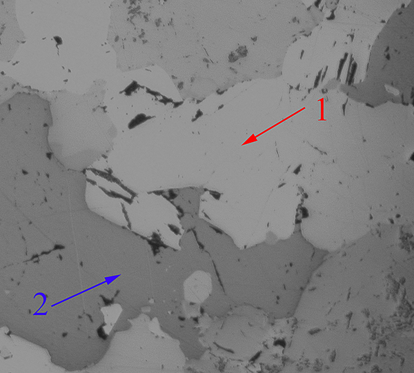
Spot 1 is a dark crystal with high refraction index. Raman spectrum above indicates Eckermanite. |
 |
 Spot 2 is a violet fluorite crystal. |
 Crystal 3 is Nepheline ( low refraction index as seen on single polarized refection image) On the top of this image, the very low index fluorite can be seen.
|
 |
 The yellow crystal (spot 4) is probably the Rosenbuschite. (or Rinkite). About the same spectrum is obtained on crystal 13.
|
|
|
|
|
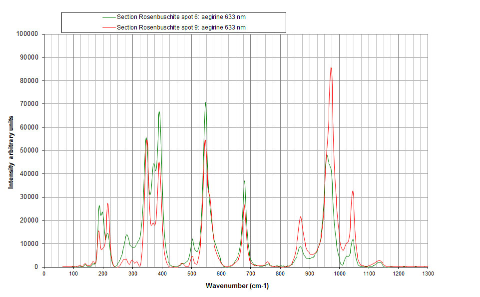
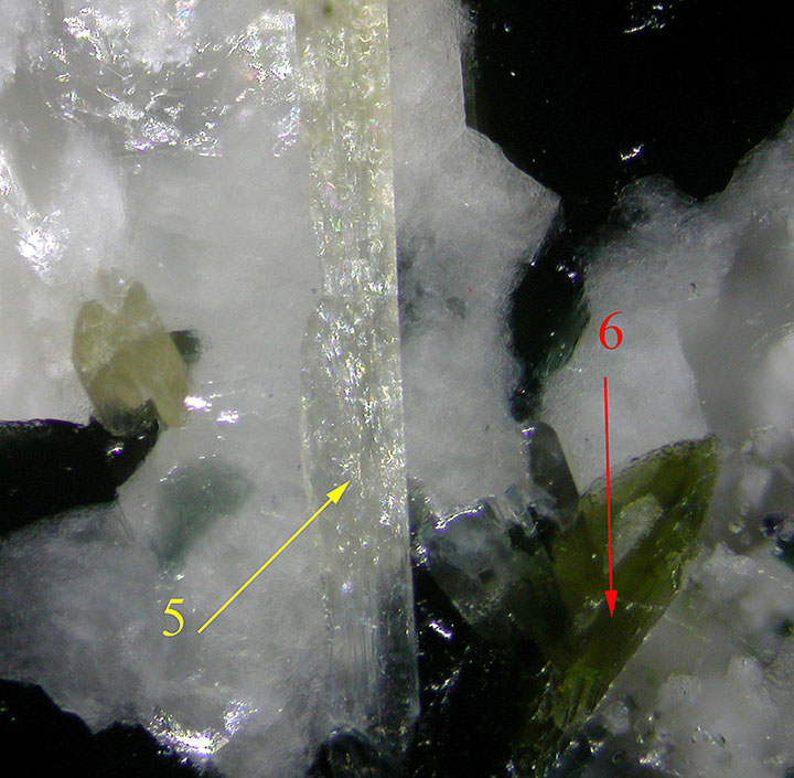 Spot 5 has a spectrum not yet fully understood: it has the same Raman spectrum as muscovite with 2 extra peaks marked in red. The green crystal 6 is Aegirine, see spectrum above.
|
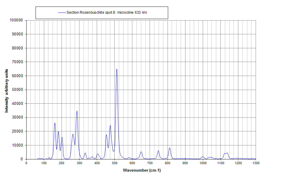
 Crystal 8 of low refraction index is microcline. |

|
 Crystal 9 is green Aegirine as can be seen on a spectrum above. |
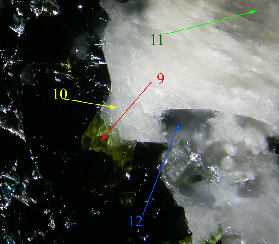 Spectrum of crystal 10 could be a mixture of 2 minerals. On the single polarized reflection image, this material seems to be altered. One of the mineral could be a mica the other could be analcime both from alteration of aegirine.
|
|
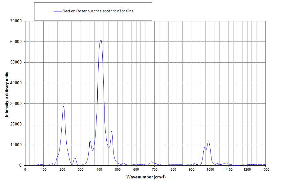
Spot 11 is Nepheline |
 The clear crystal 12 is microcline as can also be seen in thin section. |