Talc-Schist 2 / Garnet-Glaucophane
|
|
Another view of the Talc-Schist in a small
region of a thick section (~0.4 mm thickness). The pictures show a part
of a big garnet crystal ( orange in the right corner), Glaucophane
(blue-violet) and chloritoids (dark green). The chloritoids have a
strong color so that spectra have been recorded on a 30µ thin section as
illustrated on preceding page.
|
|
|
|
 |
|
|
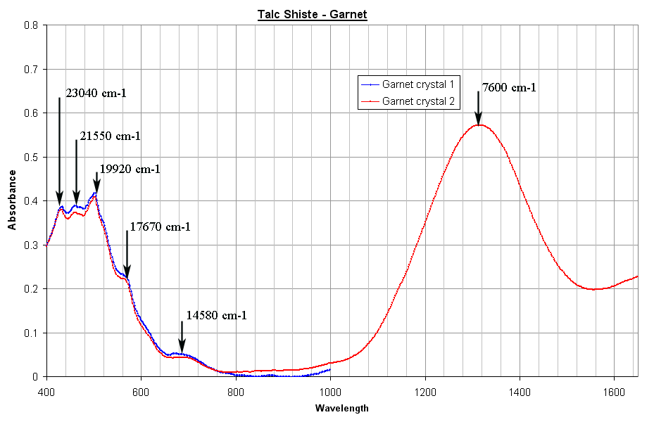
Figure above illustrates the spectrum of the garnet crystal. The garnet belonging to the cubic symmetry, its spectra do not depend upon the polarization direction of the light. The large peak at 7600 cm-1 is a crystal field transition of Fe2+ ion in a distorted 8-fold cubic site. As the symmetry of this site is no more perfectly cubic, the Fe2+ levels are further split and so produce transitions at higher wavelengths. As the wavelength range of my spectrometer is limited due to detector, only the rise of the second band at 1650 nm is barely visible. Below 600 nm, small spin forbidden transitions in Fe3+ and Fe2+ are also visible. |
|
 |
|
 |
|
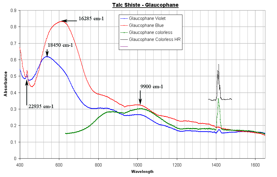
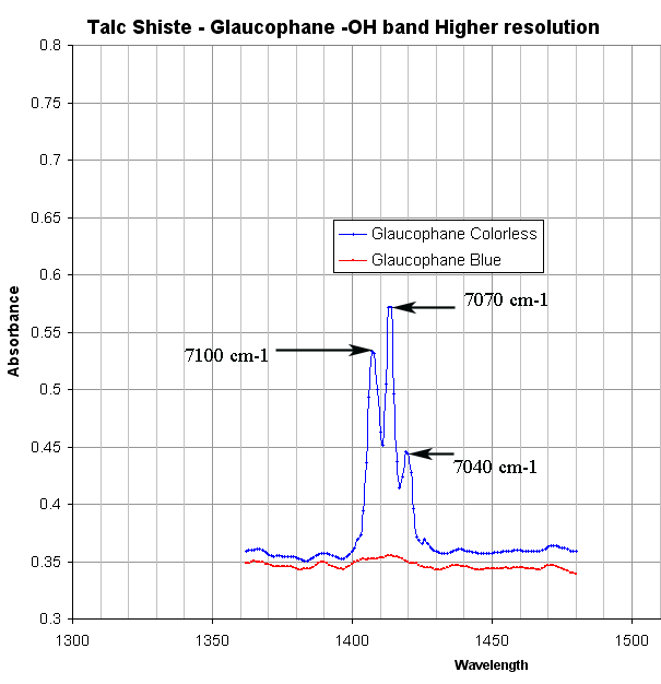
| The glaucophane visible and NIR are reproduced in the 2 graphs above. The visible spectra of glaucophane have already been presented and discussed on another rock section in the pages concerning the first microscope spectrometer. The NIR region shows 3 distinct OH harmonics. These peaks are mainly visible in the α spectrum. | |

