|
First microscope spectrometer for the measurement of the pleochroism of minerals in thin sections. |
|
|
Test of the first spectrometer with rare earth nitrates. |
|
 |
Design of a
visible - NIR spectrometer for microscope. Wavelength range : 400 to 1700 nm, transmission mode. |
 |
Microscope fiber optics spectrometer Wavelength range : 400 to 2100 nm, transmission and reflection mode. Page 1: Technical Part Page 2: Example Spectra
|
 |
Test of an Echelle Spectrograph |
 |
Connection of an ALPY 600 Spectrograph to a Microscope. |
 |
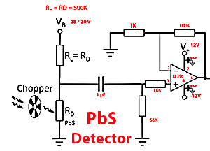
NIR Spectrometer
400 - 2500 nm Page 1: Hardware description |
|
Spectra of an epidote single crystal cut normal and parallel to crystal prism. |
|
|
Spectra of glaucophane in thin section. Page 1. Page 2. Spectra of altered glaucophane.
|
|
|
Spectra of ruby and pargasite in a zoisite-ruby section from Tanzania. |
|
|
Spectra of hornblende in thin section. Page 1. Page 2. Page 3. |
|
 |
Spectra of an Aegirine section |
 |
Pleochroism and spectra of Alexandrite. |
 |
Spectra of a Bronzite section. |
 |
Spectra of Cordierite crystals. |
 |
Spectra of a Diopside crystal. |
 |
Spectra of a Dioptase section |
 |
Spectra of an Emerald Crystal. |
 |
Spectra of an Eudialyte section. |
 |
Spectra of a Forsterite crystal. |
 |
Spectra of blue Kyanite section. |
| Spectra of Gypsum Crystals. | |
 |
Spectra of a Talc-schist with chloritoid, Garnet and Glaucophane. |
 |
Spectra of different crystal in a section of NW904 ordinary chondrite. |
 |
Spectra of olivine crystals in Seymcham Pallasite section. |
 |
Spectra of Piemontite in thin section. |
 |
Spectra of Tourmaline. |
 |
Spectra of a Pyroxenite section. |
 |
Spectra of Garnet. |
 |
Spectrum of Vivianite |


