| The Raman microscope can easily
determine the mineralogy of a granite polished slab as shown below. All
images are reflected light images with oblique illumination. |
|
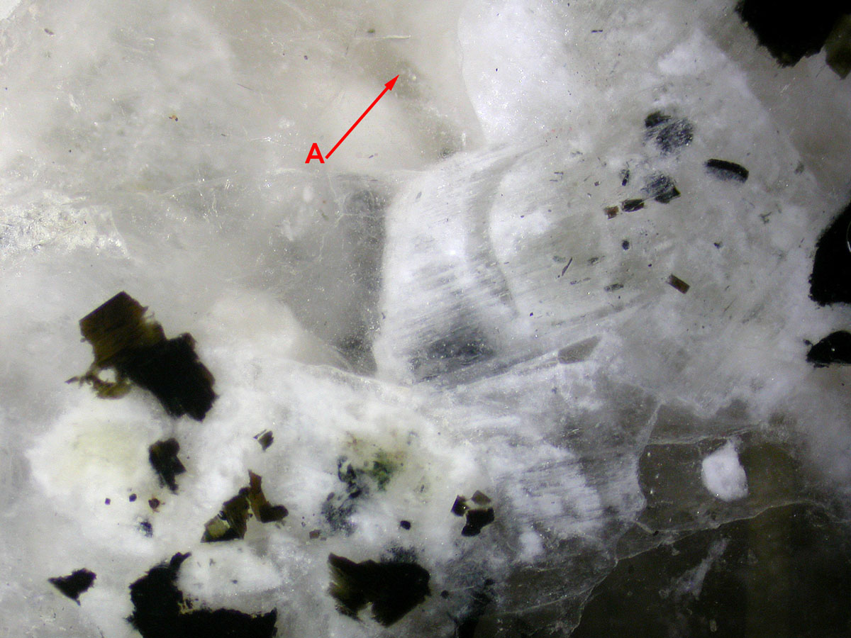
Granite spot A: Quartz |
 |
|
Optically, it was not easy to see if spot A was Quartz or Feldspar. |
|
|
 |
| The Raman spectrum shows that the
mineral of spot A is evidently Quartz. |
| |
|
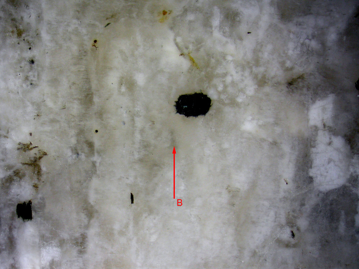
Granite spot B: Feldspar |
 |
|
At first sight on a reflected light image the difference between spot A
and B was not self evident. |
|
|
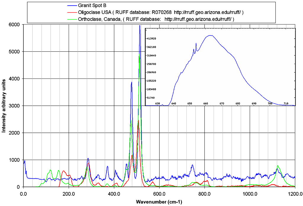 |
| The Raman spectrum of spot B indicates
the presence of a Feldspar. Notice that the fluorescence of this white
diffusing mineral is very high as can be seen on the original signal
before baseline removal. This high fluorescence and the low Raman
intensity are responsible for the
low signal to noise of this spectrum. |
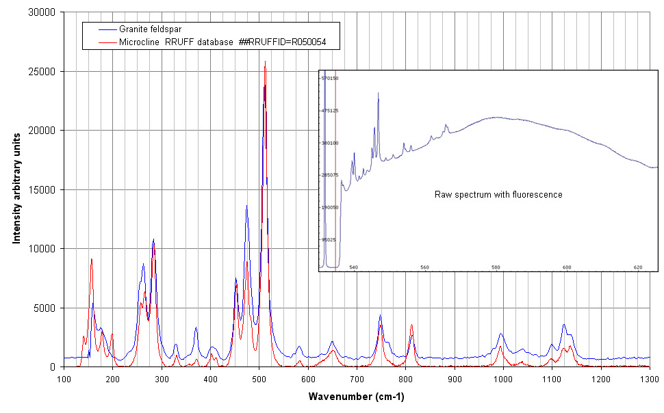 |
| Raman spectrum of a feldspar crystal in
the same granite section with new spectrograph and laser 532 nm.
Fluorescence is still present (see raw spectrum) but the signal to noise
is much higher. |
|
Granite spot C: Mica + Quartz crystal |
 |
|
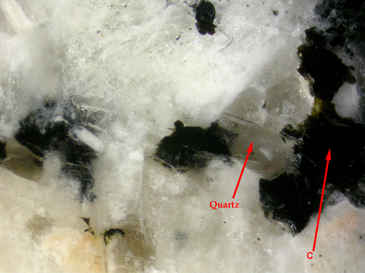
Two crystals have been targeted : a transparent one which seems to be
Quartz and a dark one (spot C) presumably a mica Biotite. |
|
|
 |
|
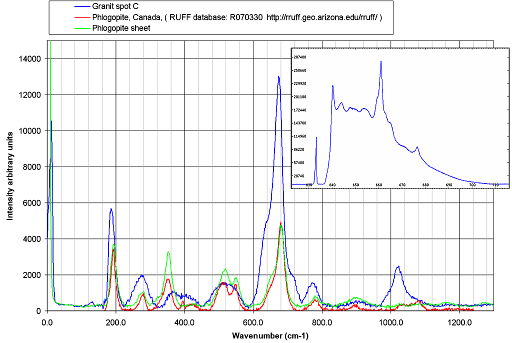
Raman spectrum shows that spot C is a mica of the Phlogopite family, the
dark color indicates Biotite as expected. |
|
|
 |
|
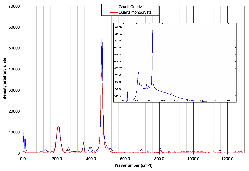
Raman spectrum confirms the structure of the big transparent crystal
above as Quartz. |


