|
Rutile Goethite Calcite |
 |
|
Spot E is a brownish material abundant in this rock section while
spot D is a dark red-brown crystal of a minor mineral. A higher
magnification image is reproduced below. |
|
|
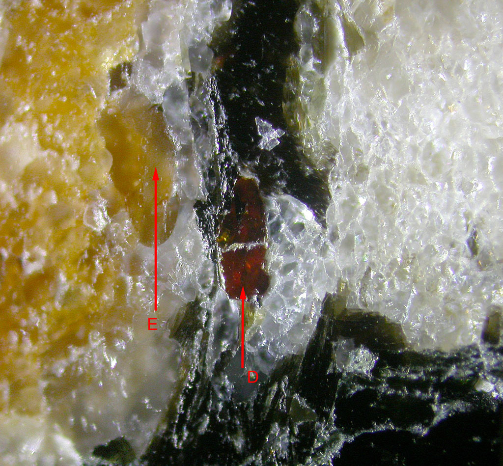 |
| Higher magnification image showing
the color difference between both crystals D and E. |
| |
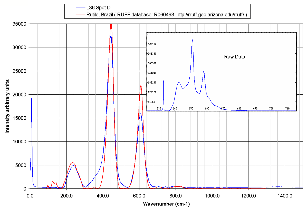 |
| Raman spectrum of spot D
indicates clearly the presence of Rutile. Rutile and Ilmenite
detected previously confirm the presence of titanium in this rock. |
| |
 |
| The structure of the brownish material (
spot E) is elucidated by the Raman spectrum: it is made up of Calcite
and Goethite. The detailed structure of this two minerals
assemblage is illustrated in the thin section images below. |
| |
 |
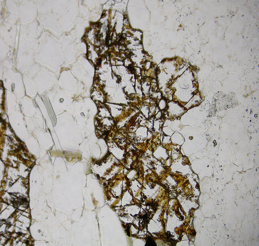
| Enlarged (10X objective) thin section image
showing a high interference color calcite material (pink or green
depending on polarization vector) with embedded goethite veins.
Depending on the precise position of the laser spot, calcite alone or a
mixed spectrum of calcite and goethite can be obtained. The
section shows also the low birefringence albite crystals and some highly
colored muscovite. |
| |
 |
| Same view without analyzer inserted. The
Goethite network is clearly visible. |
| |
 |
| Another part of the thin section
with the green-pink carbonate with goethite inserted in the fractures of
the grains. |