Mangano-Calcite Fluorescence
See the Raman spectra of this section
|
|
|
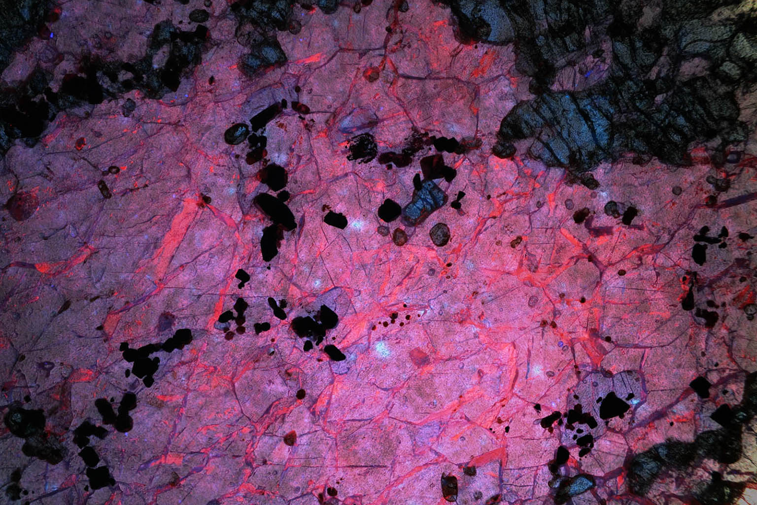
Figure 1: transmission fluorescence image of a section of mangano-calcite and forsterite from Sweden. The mangano-calcite is fluorescent.
|
 |
|
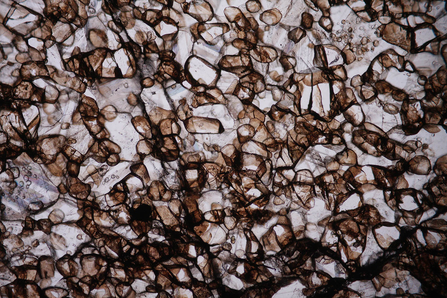
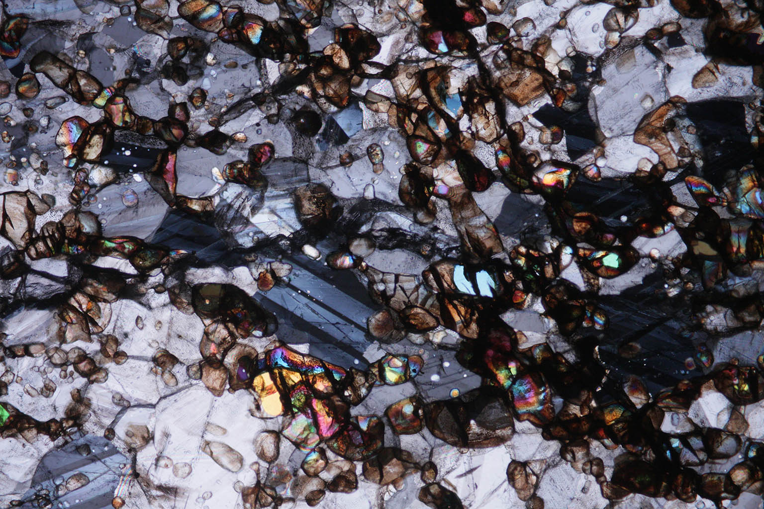
Figure 2: same section in plane polarized light. The dark mineral is hausmanite.
|
 |
|
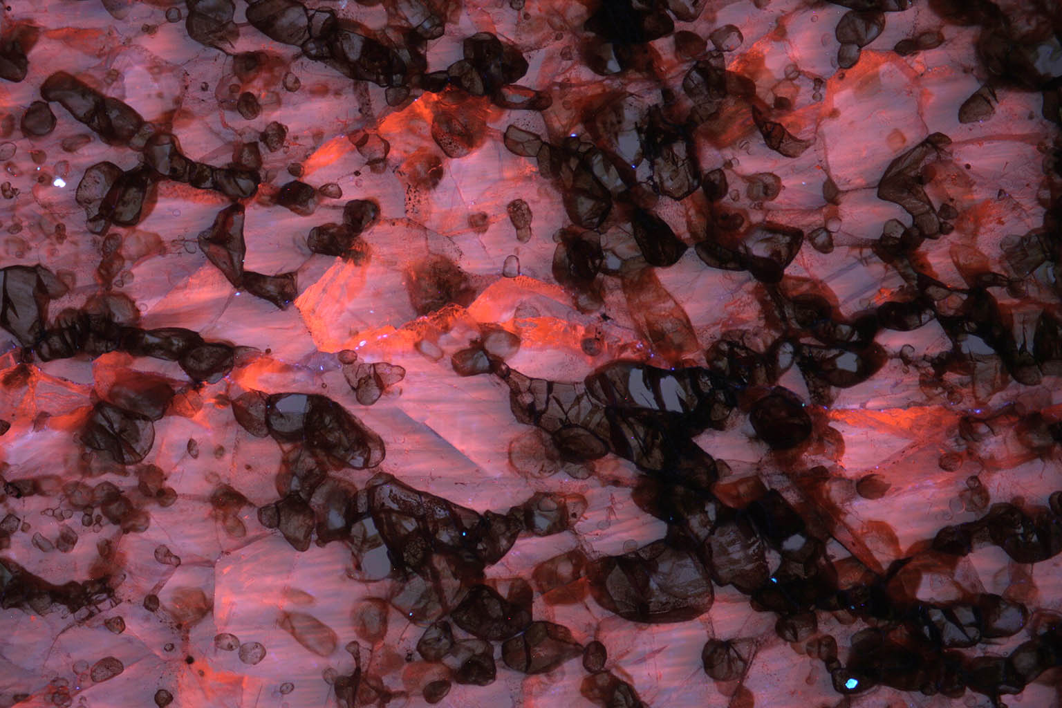
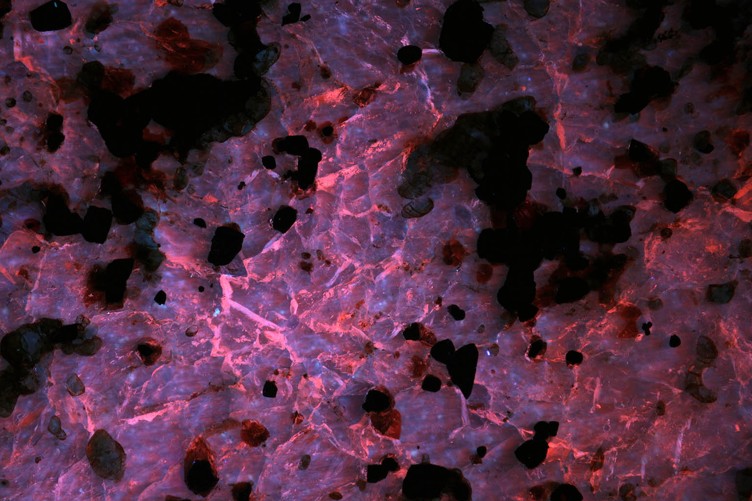
Figure 3: same mangano-calcite sample fluorescence in reflection.
|
 |
|
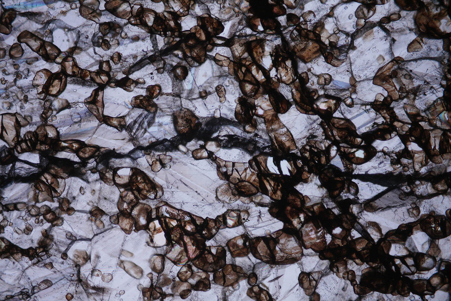
Figure 4: transmission plane polarized view.
|
 |
|
Figure 5: crossed polars view. The bright interference colors come from forsterite.
|
 |
|
Figure 6: another sample from the same place. Transmission fluorescence.
|
 |
|
Figure 7: same sample viewed in reflection fluorescence. The fluorescence seems to originate from the rims of the crystals.
|
 |
|
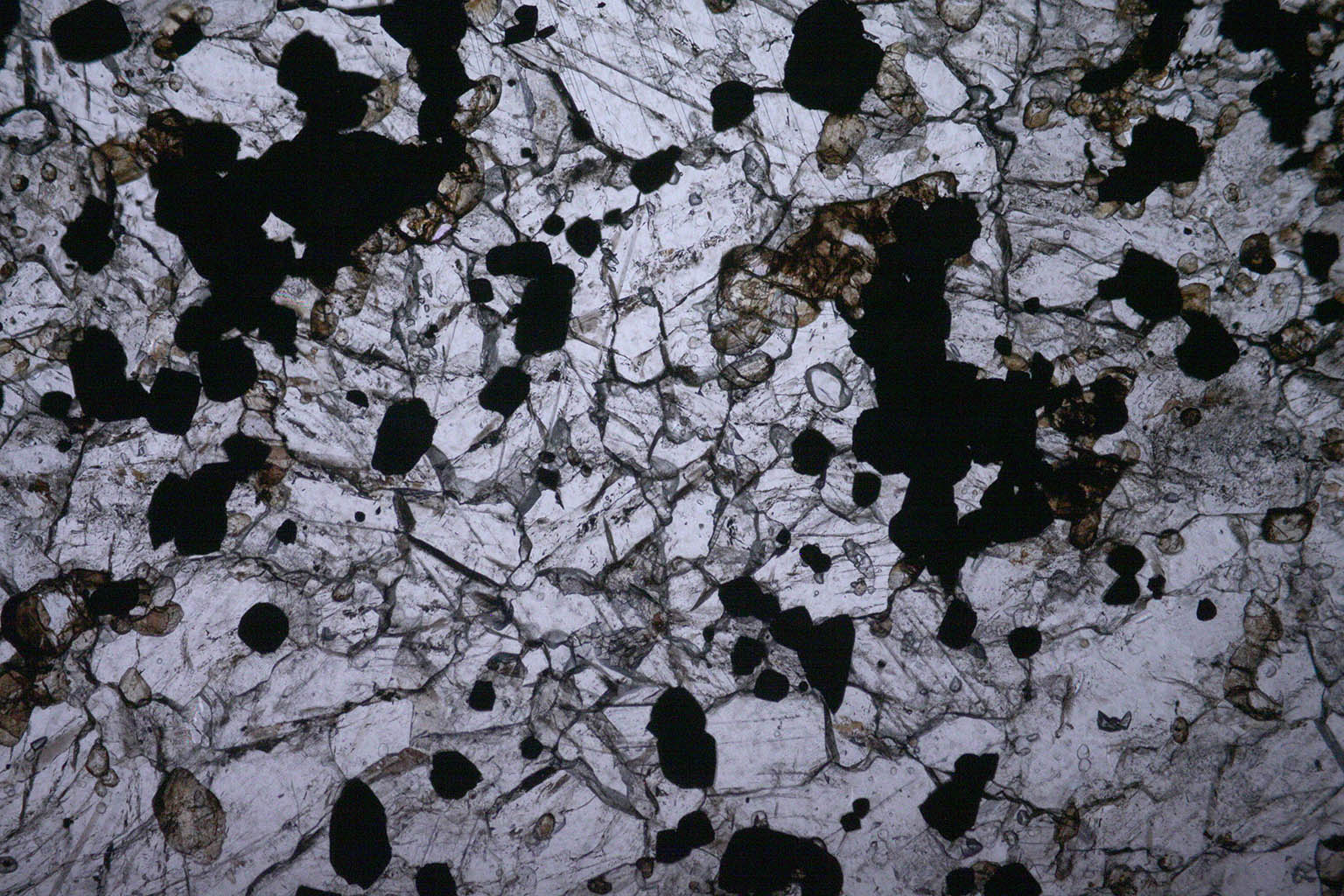
Figure 8: same section in transmission plane polarized view. A higher amount of opaque hausmanite and less forsterite can be seen compared to the first sample.
|
|
|
|
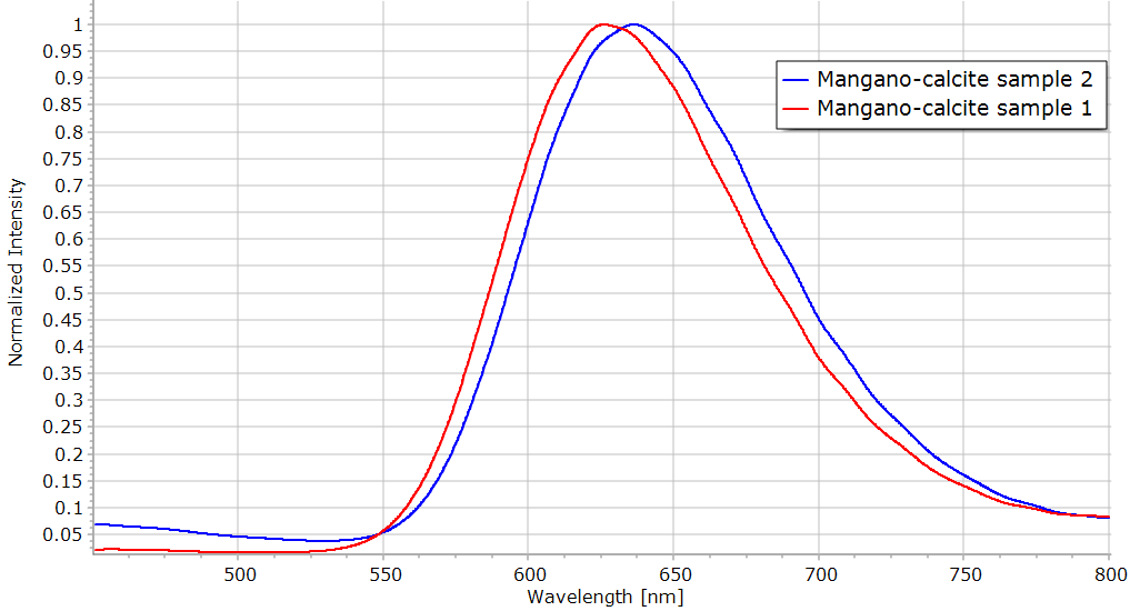
Figure 9: fluorescence spectra of the two samples recorded with the Alpy 600.
|