Conoscopy of Biaxial Minerals (3).
Figure normal to one optic axis.
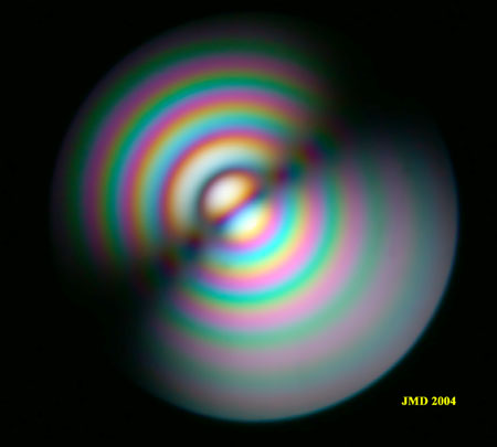
The optic normal figures below were photographed from a thick Danburite crystal whose prism faces are nearly perpendicular to the optic axes. The angle between optic axes is approximately 90°. The dark isogyre is approximately a straight line due to the high angle between optic axes. The isochromes are approximately circles because, due to the high 2V angle, the melatopes are located far from each other and the lemniscates joining both poles of the optic axes are located far beyond the field of view. In white light (figure 1), only a few rings can be seen due to the fact that high orders are quickly reached when moving off axis. Figure 2 photographed in monochromatic light exhibits many rings up to 17th order.
|
Figure 1. Thick Danburite crystal. White light. |
Figure 2. Monochromatic light, 578 nm. |


In figure 3, the counter rotation of the isogyre with the stage can be examined. Point X rotates clockwise with the microscope stage while the dark isogyre rotates counterclockwise.
|
Figure 3a. Rotation of the isogyres. |
Figure 3b. |
Figure 3c |


Below, a thin danburite crystal cut perpendicularly to the optic axis has been photographed. The number of rings of the isochromes is of course much reduced. The addition of two accessory plates is also illustrated. As the angle between the optic axes is close to 90°, it is not possible to determine from the curvature of the isogyres what is the direction of the acute bisectrix so the accessory plates cannot be used in this case to determine the optic sign.
|
Figure 4. Thin Danburite crystal cut normal to optic axis. |
Figure 5. Same crystal with addition of a wave plate. |
|
Figure 6. Same crystal with insertion of a quarter wave plate. |


Next page [Conoscopy main page] [Home]