Alexandrite spectra.
![]()
|
|
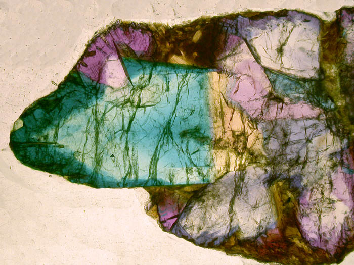
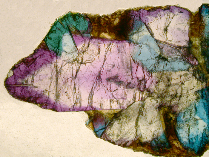
Alexandrite section from Bahia, Brazil (~0.5mm
thickness). This picture and the following illustrate the beautiful
pleochroism from green to yellow to pink of the Alexandrite.
The spectra below were recorded on the green-pink and yellow-pink parts of the crystal changing the orientaion of the polarizer by 90°. The presence of Cr3+ ions in the chrysoberyl structure gives the bimodal absorption feature responsible for the color and the pleochroism. |
|
|
|
|
|
|
|
|
This enlargement of the region between 600 and 700 nm shows low intensities spin forbidden transition of the Cr3+ ion. The alexandrite spectrum is quite comparable to Ruby as presented previously. |