Raman spectra of Beryl and Emerald
![]() Raman Main Page
New spectrograph spectra.
Raman Main Page
New spectrograph spectra.
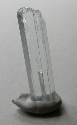 |
Small crystal of very pure Beryl,
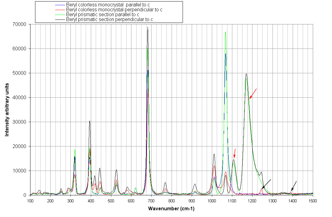
colorless. The spectra red and blue in the figure below have been
recorded with this crystal. Another beryl section, slightly colored
(spectra green and black) has also be examined. Spectra of both crystals
exhibits the features of Beryl material as found in the literature
except for the two bands marked with a red arrows which appear only on
the prismatic section. It is again believed that these peaks are not
Raman vibrations but fluorescence of Cr3+ ions. The two small peaks indicated by black arrows in the figure could be attributed to CO2 molecules which can be present in the rings of the Beryl crystal ( CO2 Fermi doublet). |
 |
|
 |
|
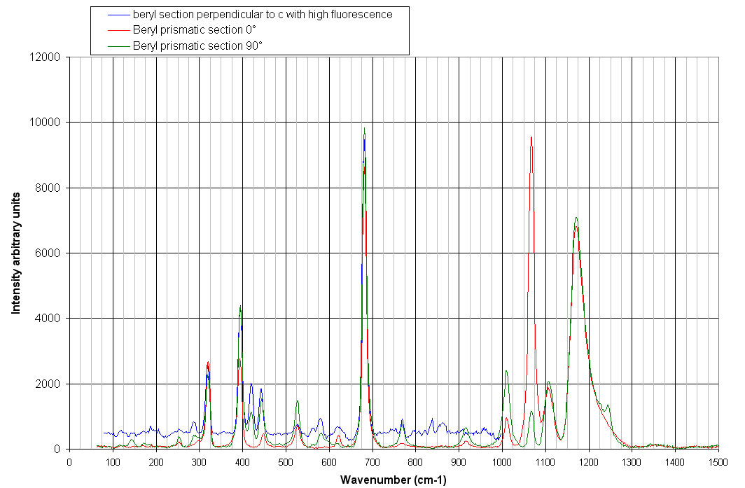
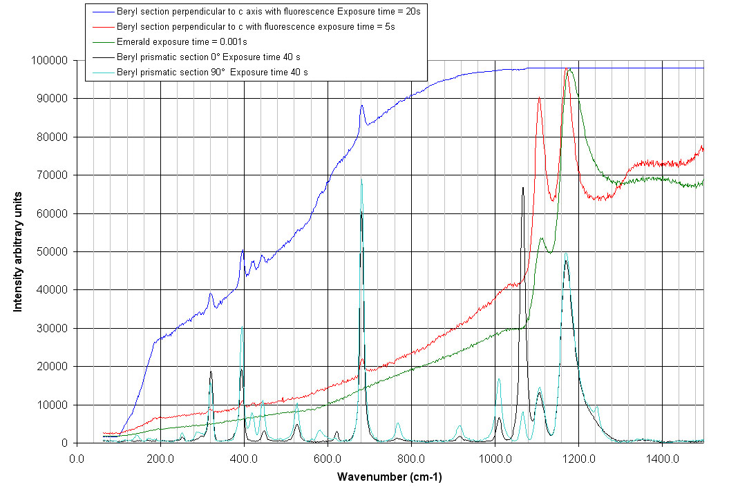
| Some samples exhibit a high fluorescence. The sample with the blue spectrum in the figure above is such a sample. The background corrected spectrum is much more noisy but the main features below 1000 cm-1 can be attributed to Beryl. | |
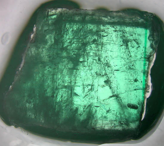 |
To look further at the interpretation of the 2 bands at 1105 and 1175 cm-1 , the raw spectra of the high fluorescence Beryl and an Emerald crystal have been superimposed on the background corrected spectra shown above. Note the great difference of the exposure times to get a signal in the range of the CCD. The blue curve below is the raw signal of the blue spectrum reported above. Note that the CCD is completely saturated above 1000 cm-1 . If the integration time for this Beryl is decreased (red curve), two high intensity peaks appear around 1105 and 1175 cm-1 . The Raman spectrum below 1000 cm-1 is barely visible for this integration time. In order to get the signal for an Emerald crystal in the range of the CCD, a very short integration time (1 ms) must be used. The same two peaks are already present in the spectrum for integration time with very high intensities. The color of Emerald is due to impurities of chromium 3+ which give here a high fluorescence. If a green laser is used for Raman excitation, the position of the chromium peak do not move (in nanometers) and thus this fluorescence should appear well above the Raman spectrum. Chromium fluorescence is a disadvantage of He-Ne laser for Raman spectroscopy.
|
 |
|
 |
|
|
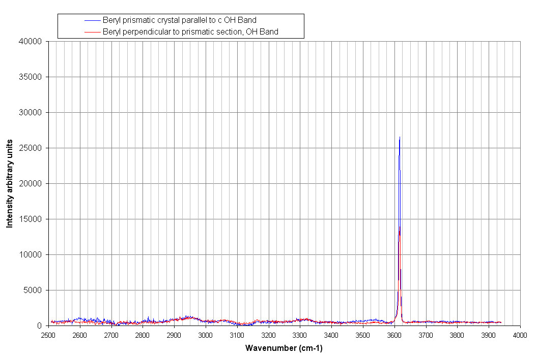
Water molecules can also be present inside the ring channels of Beryl as demonstrated by the spectrum above on the stretching OH band at 3615 cm-1 . |
|