Glaucophane Prasinite from Viu Italy. Page 5.
Muscovite |
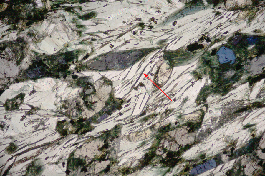
| The muscovite, transparent on a transmission image is a major mineral in this rock sample. It appears with brilliant interference colors throughout the thin section. |
 |
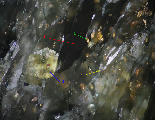
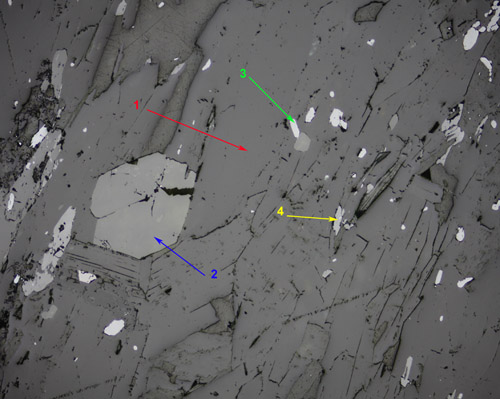
| On the reflection image (left below), the muscovite appears dark due to the light absorption of the minerals deeper in the section (glaucophane) |
 |
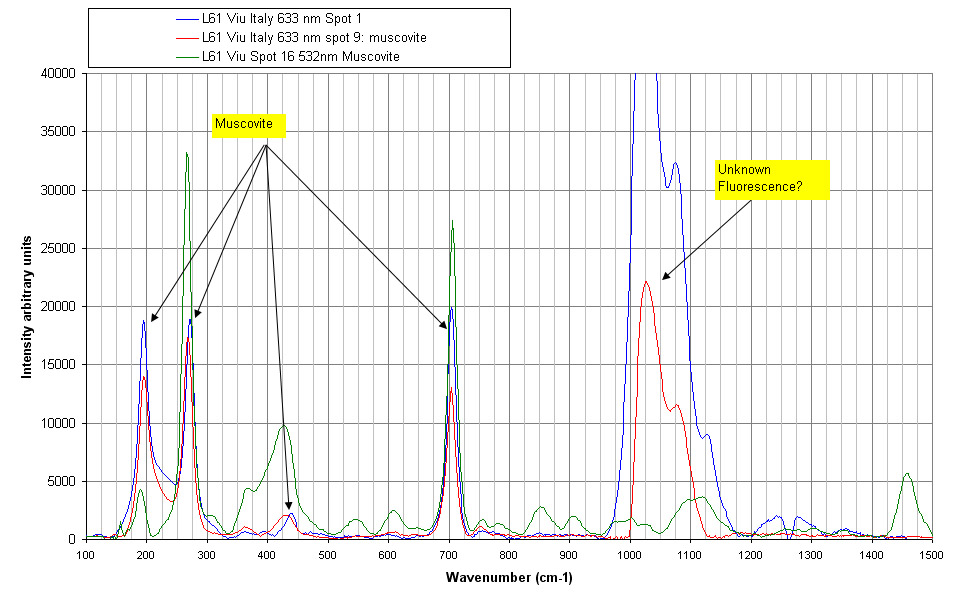
| Muscovite Raman spectra at 532 nm. |
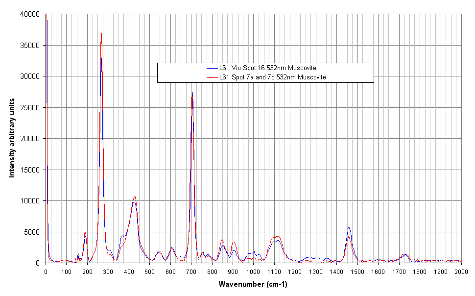 |
| With the red laser, spurious lines appear in the Raman spectrum. Those lines are absent in spectra got with green laser. Fluorescence? To be further investigated! |
 |
Hematite |
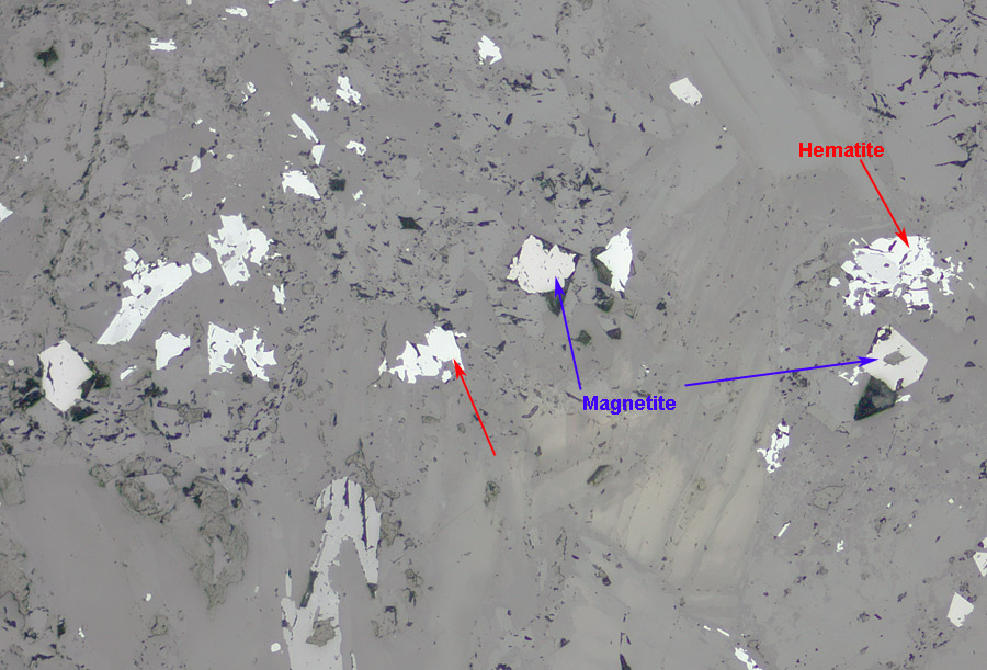 |
| Picture of the opaque minerals. Magnetite and hematite have been identified in this section. Hematite has a higher reflection coefficient. |
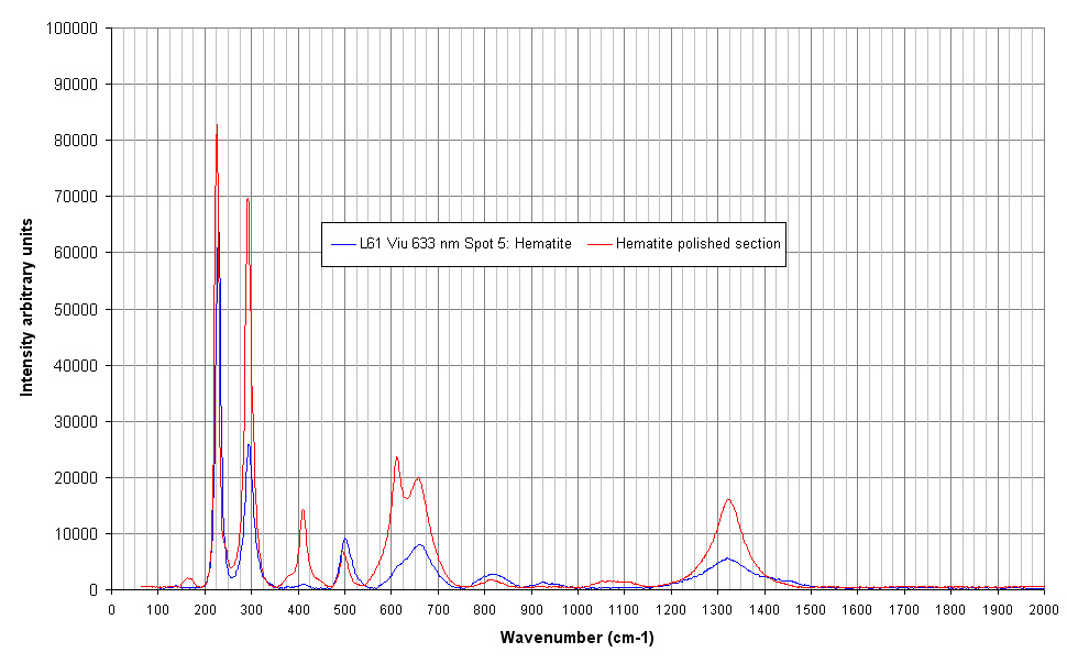
| Hematite Raman spectrum compared with a pure hematite sample. |
 |
Magnetite |
|
|
| Magnetite is also identified by Raman. The intensity of the laser should be decreased to see the magnetite spectrum. If light intensity is too high, the magnetite is oxidized and an hematite spectrum is obtained. |
