Raman Sodalite from Norway.
Fluorescence page of this section.
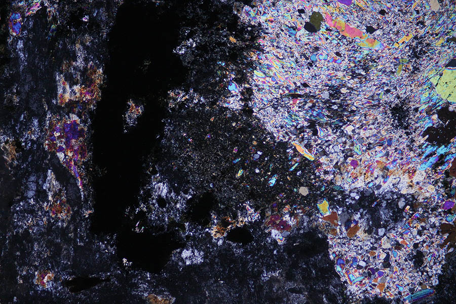
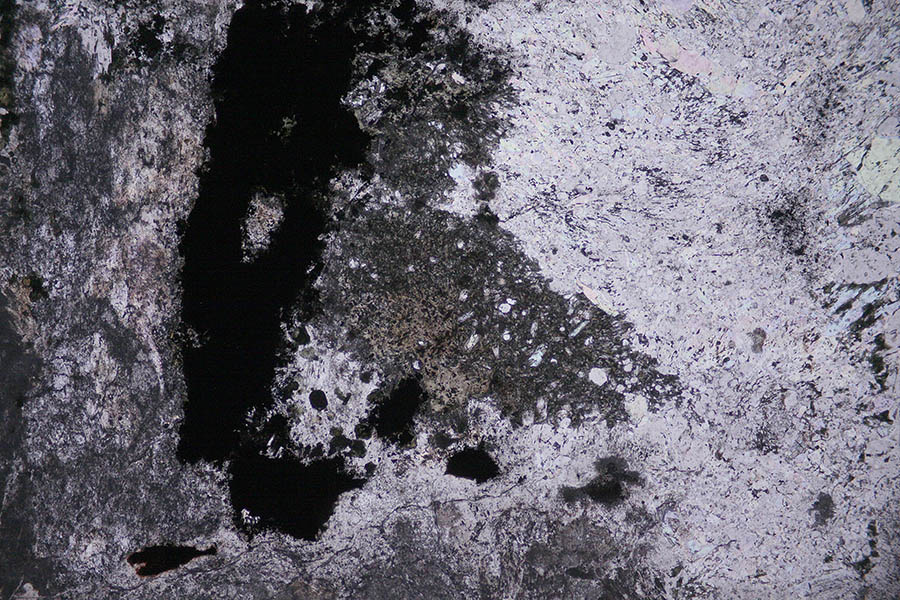
| Raman study of a highly altered by weathering sodalite section from Narvick, Norway. Due to the weathering of most of the minerals of this rock, the section does not give nice pictures. Sodalite is accompanied by sodium feldspar, cancrinite and some zeolites sometimes difficult to identify formally. Some chlorite family minerals are also suspected. Minor minerals are ilmenite, molybdenite... This section is about 100 µ thick to avoid Raman spectrum of the glue so the crossed polars views (LPA) give uncommon polarization colors.
|
|
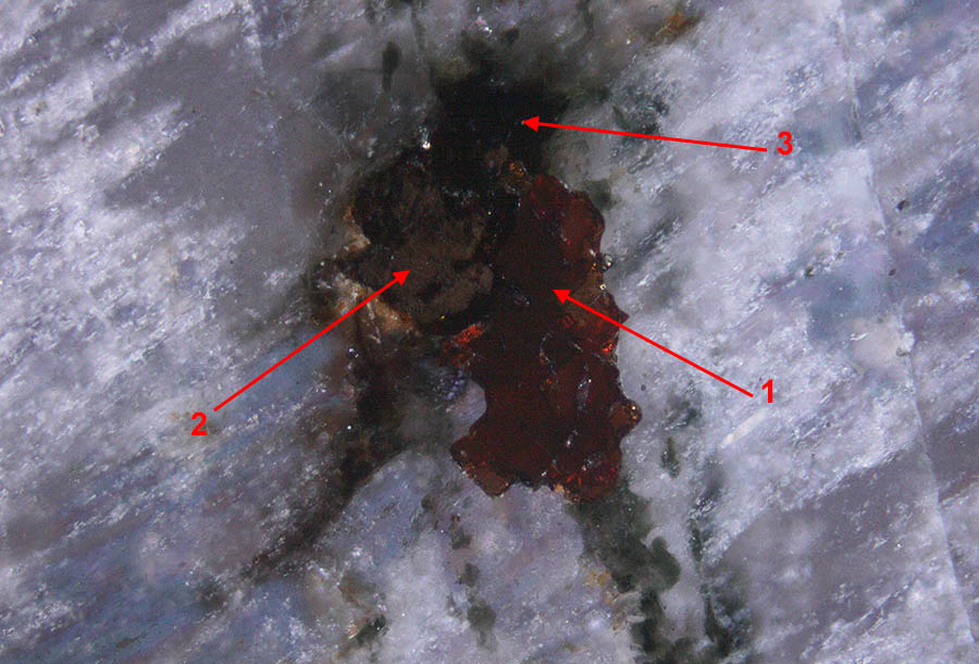
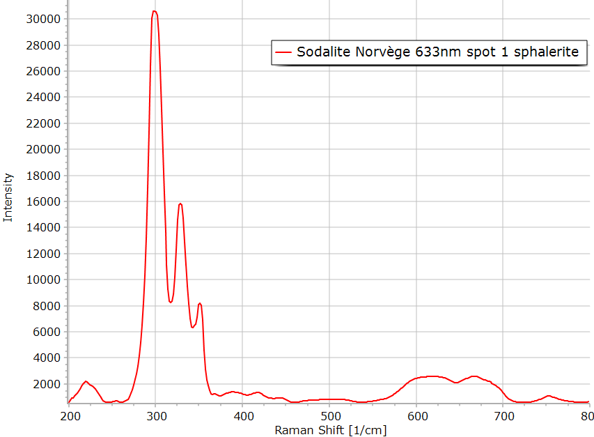
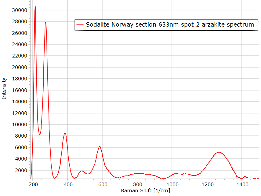
Reflection view of small opaque inclusions in the rock. Very bright zone 2 has brown internal reflection in the double polarized image on the right. Area 1 is the mineral sphalerite as can be seen on the spectrum below. The clear yellowish mineral in zone 2 could be arzakite a mercury chloro-sulfide as indicated by the Raman spectrum below. This determination of arzakite is not sure because the color seems to be too much yellow.
In this doubly polarized reflection image, the sphalerite exhibits red brown internal reflections.
|
|
|
|
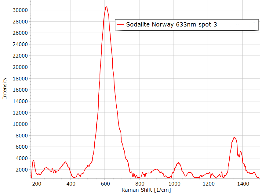
Mineral in area 3 has not been identified with certainty. The Raman spectrum is close to pyrochlore but it is difficult to be sure in this high fluorescence section. |
|
Another view of the minerals present in zones 1 and 2. The image on the right confirms the color of the minerals with the internal reflections. |
|
Transmission image, crossed polars. |
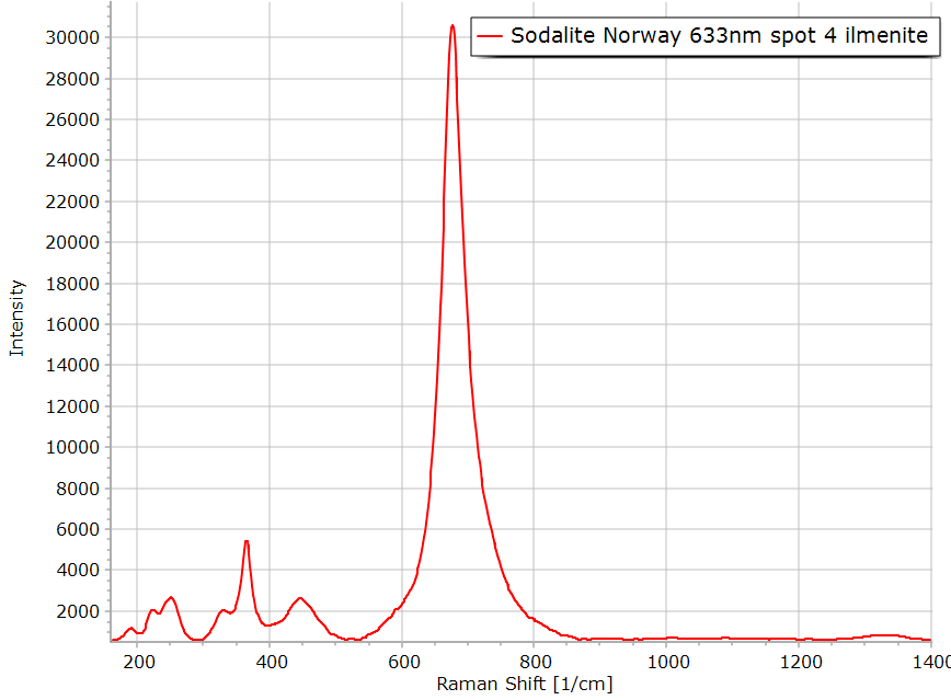
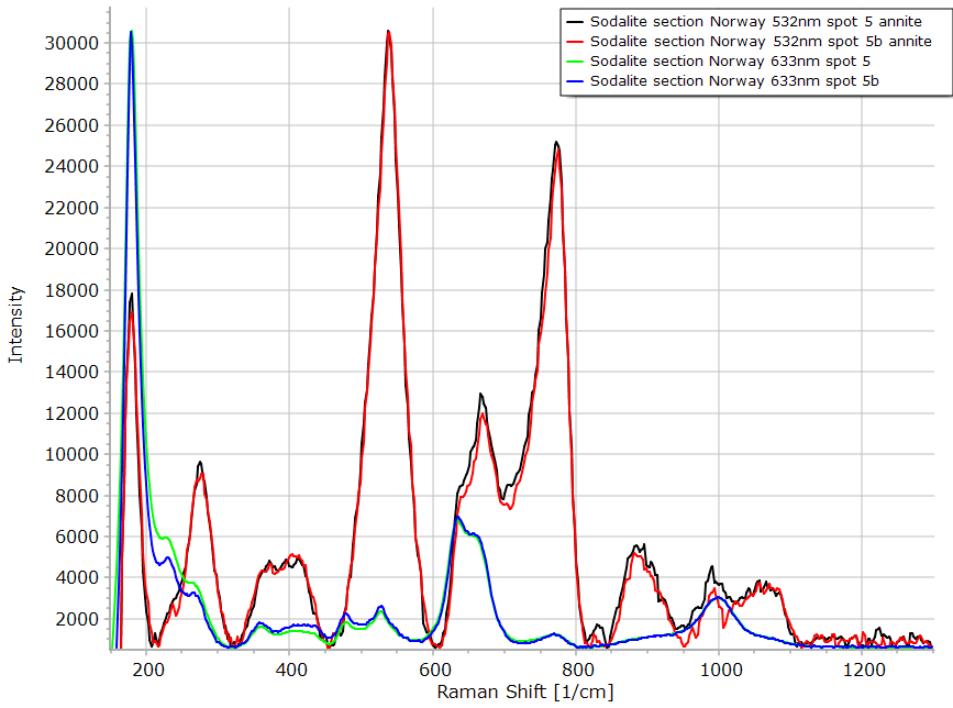
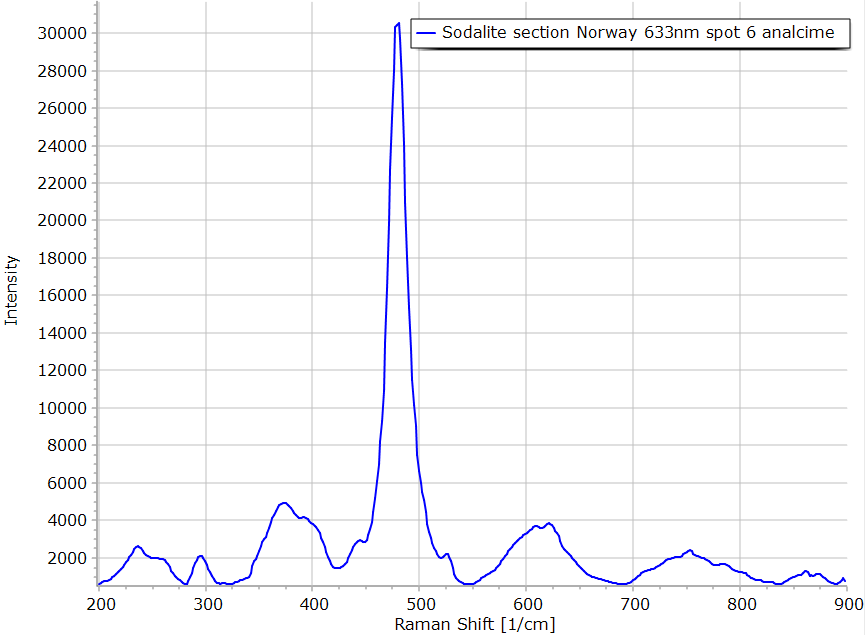
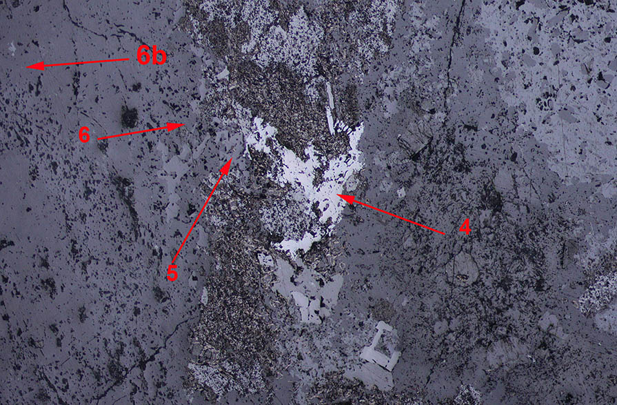 Reflection LPNA image. The figure above shows 3 different minerals in spots 4, 5 and 6. In zone 4, the higher reflection coefficient mineral is ilmenite. In area 5, Raman indicate the presence of the mica annite. The spectra of this location with the 632.8 and 532 nm laser are very different without explanation up to now. In zone 6 and 6b, we have found the zeolithe analcime.
|
|
Transmission plane polarized image
|
|
|
|
|
|
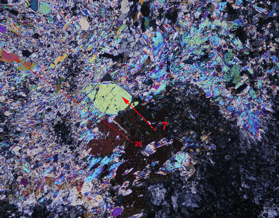
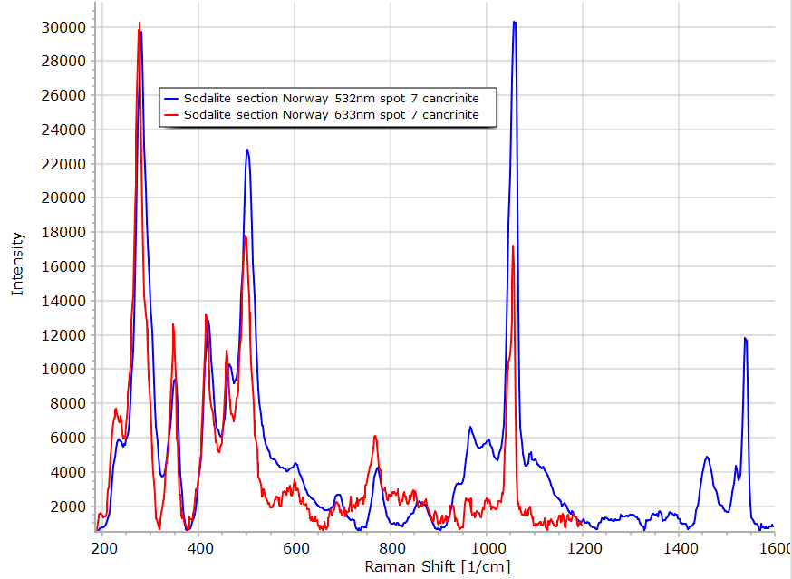
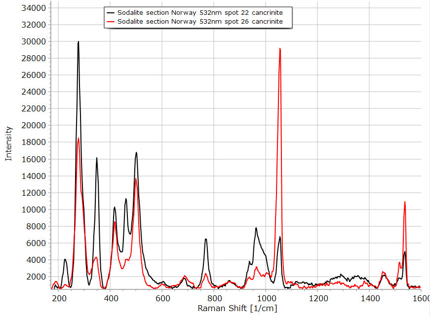
The birefringent crystal seen in zone 7 is cancrinite. The other
minerals around it are also cancrinite as illustrated by the spectrum of
area 26 below.
|
|
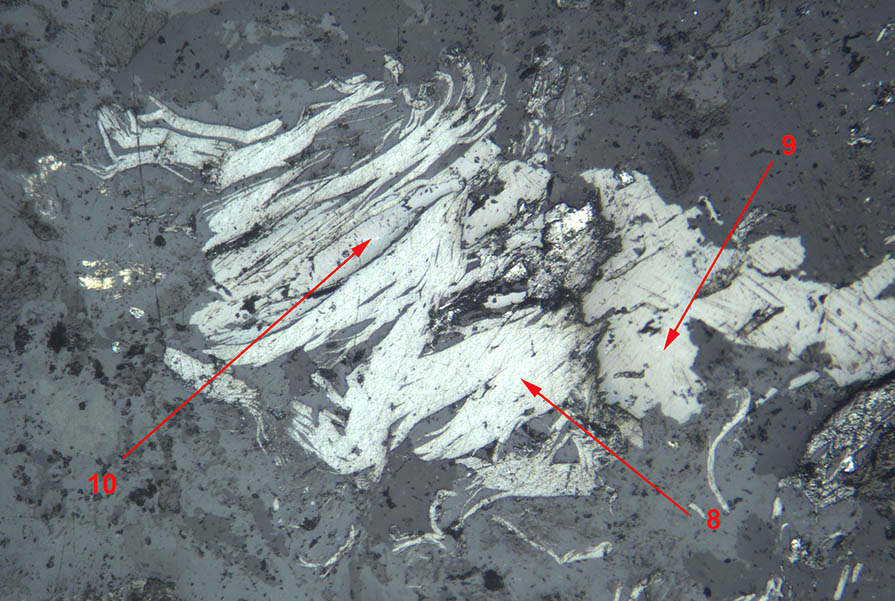
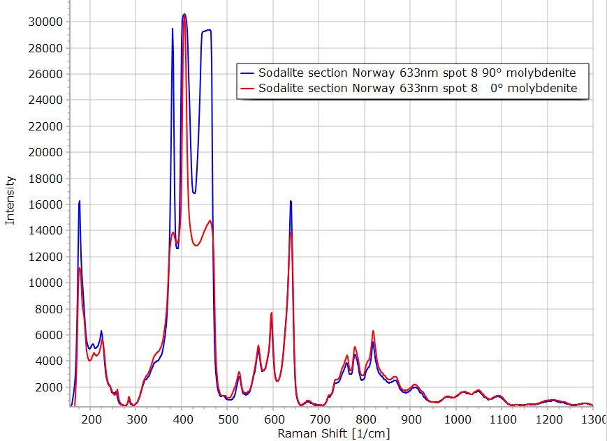
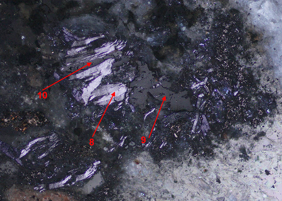
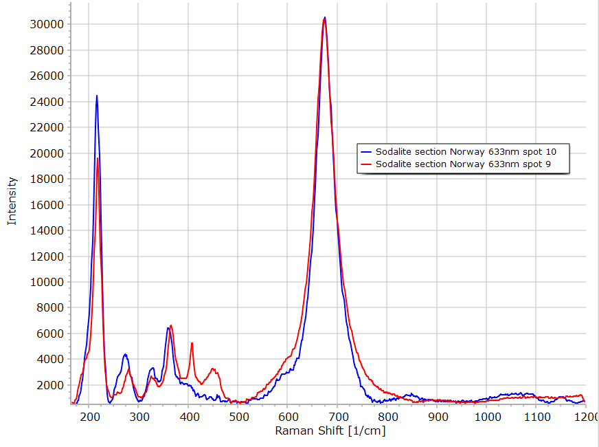
Another opaque inclusion in this section. Single polarized reflection view above. Area 8 is a high bireflectance material highly anisotropic as can be seen in double polarized image below. The Raman spectroscopy identifies this mineral as molybdenite. Material in areas 9 and 10 could be ilmenite as suggested by the anisotropy in the image below and the Raman spectra although some additional peaks are present.
|
|
|
|
|
|
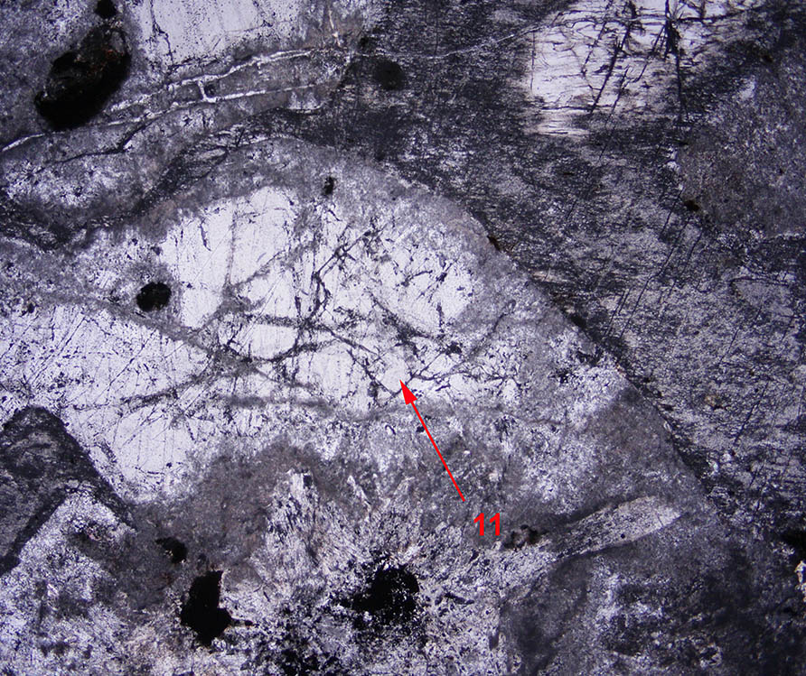
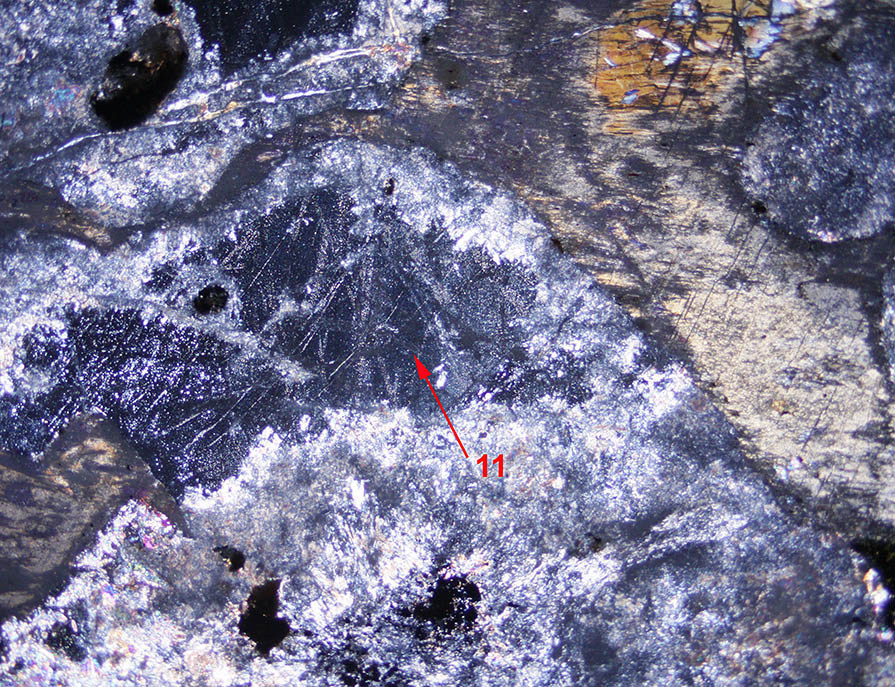
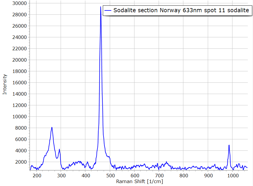
The cubic crystal in area 11 is the sodalite. Notice the alteration in the lower part of the image identified by fine grained anisotropic material. |
|
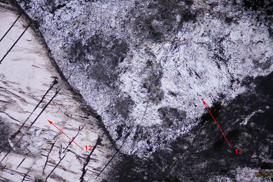
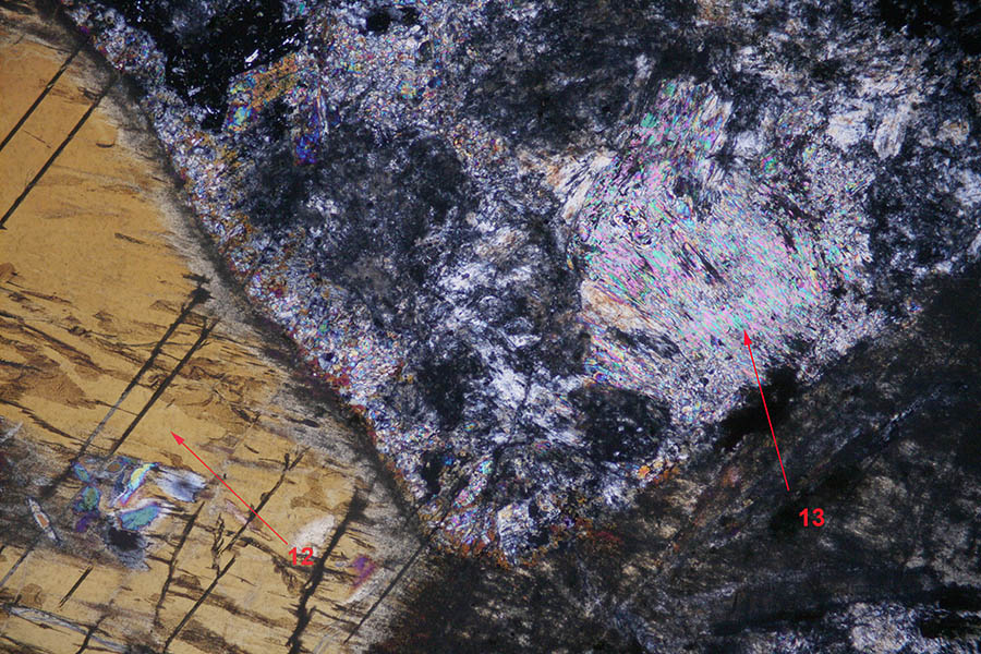
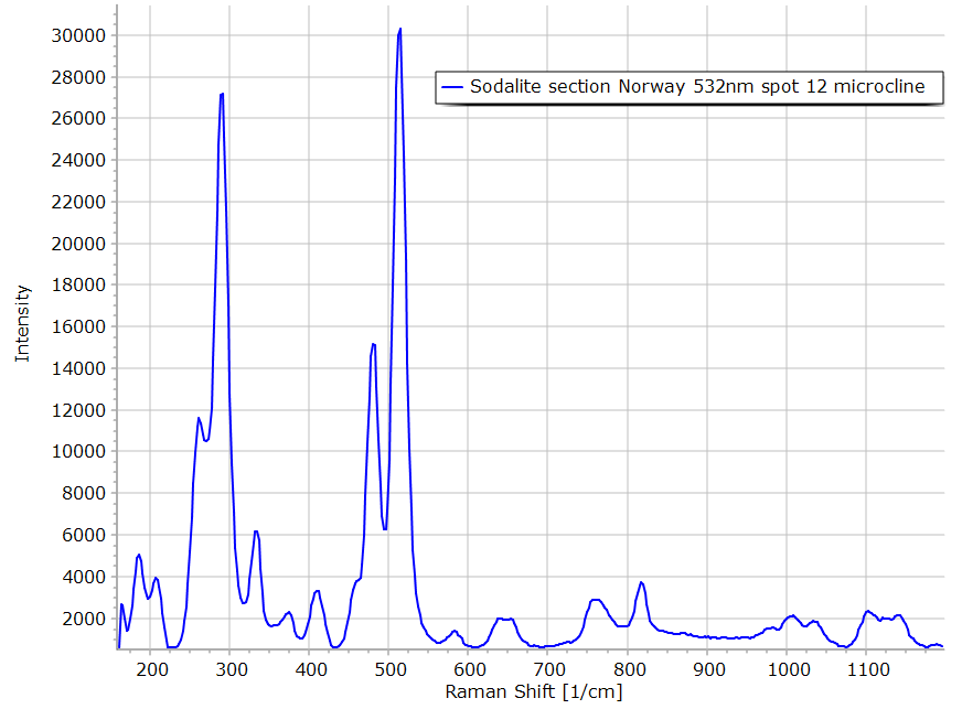
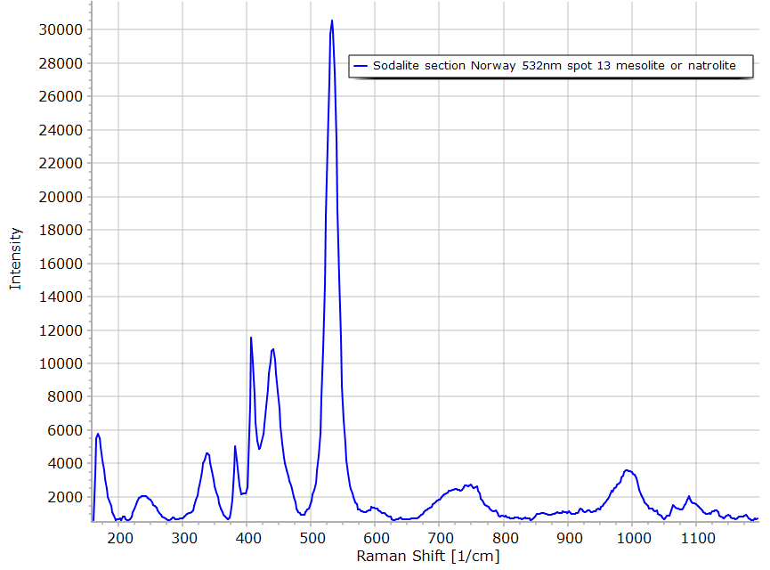
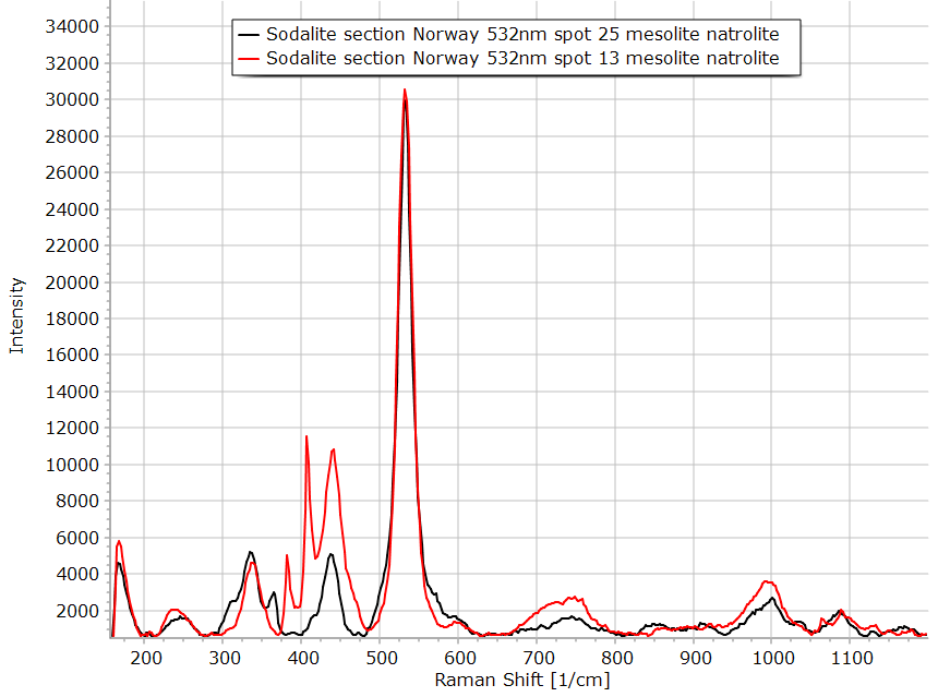
On the left, area 12, a big crystal of microcline has been found. On the right, a big crystal highly altered has been analyzed by Raman. The spectrum indicates the presence of a zeolite, it is closer to the mesolite spectrum but could also be natrolite. See below on the right.
|
|
|
|
|
|
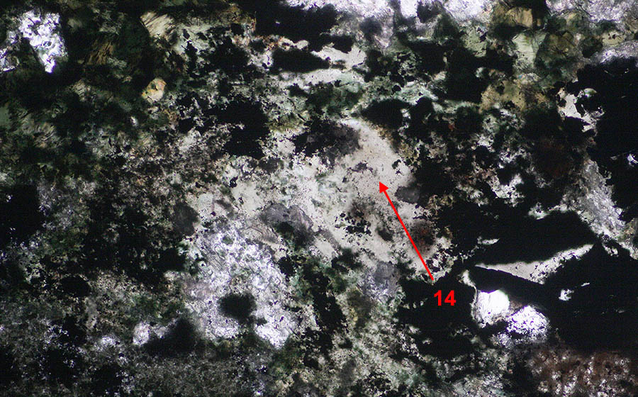
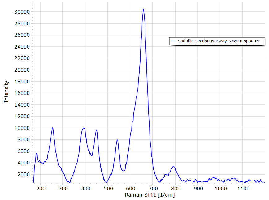
The clear brownish material found in area 14 has not been identified with the help of the Raman spectrum on the right.
|
|
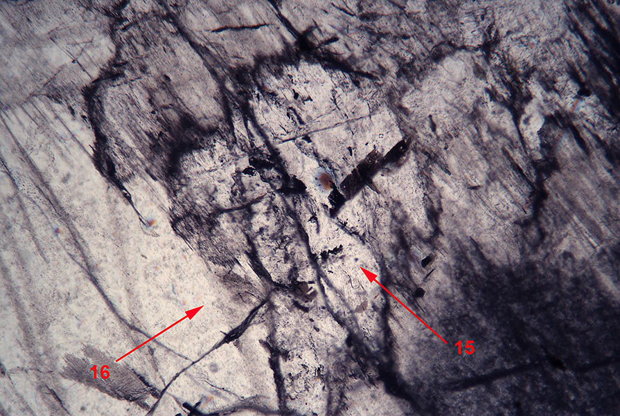
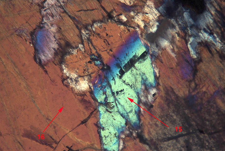
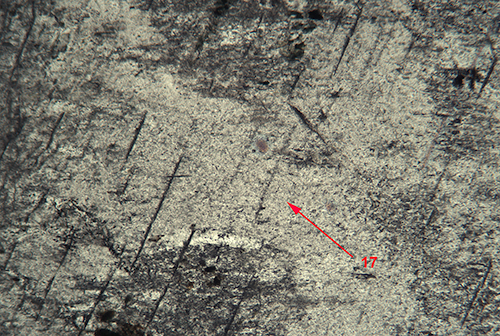
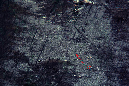
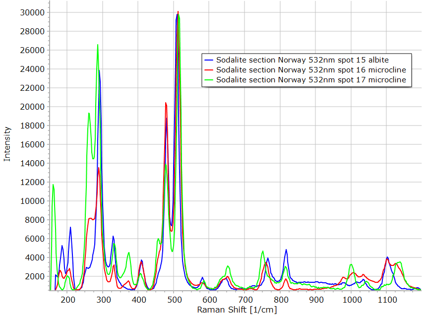
On the top 2 figures (areas 15, 16 and 17), we find the sodium feldspars microcline and albite. |
|
|
|
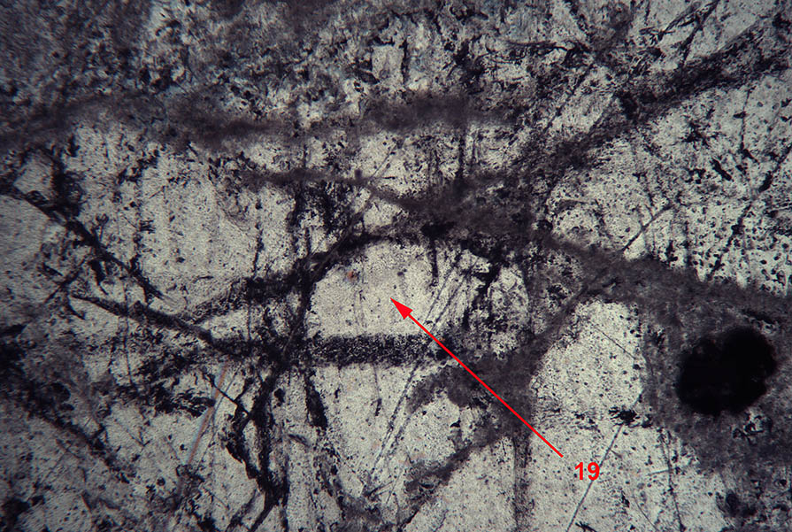
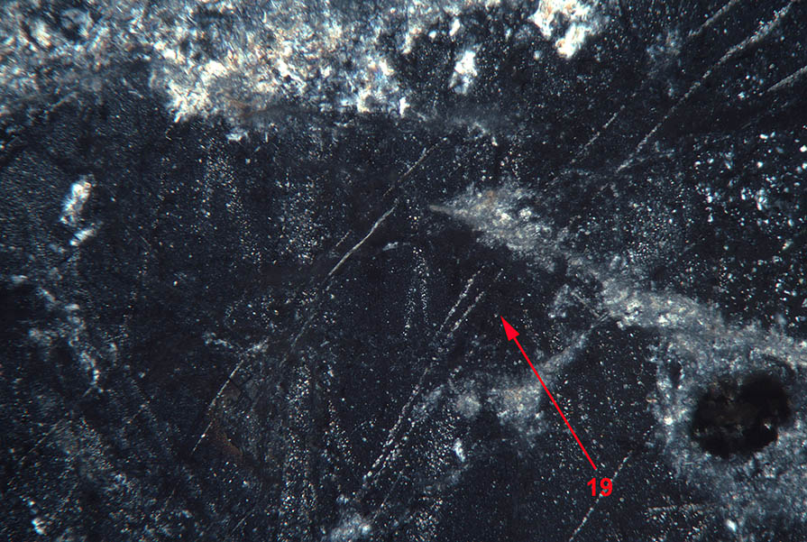
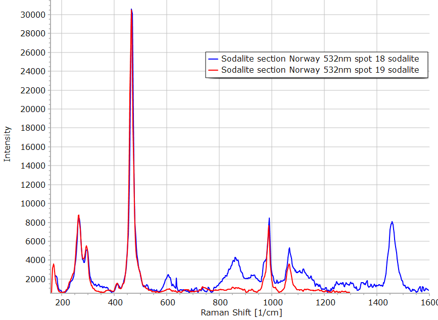
The cubic material present on the crystal Nr 19 is again sodalite. Fine grains alteration material appear bright on the LPA image. |
|
|
|
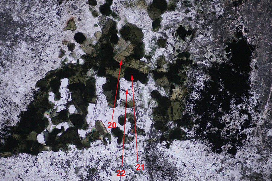
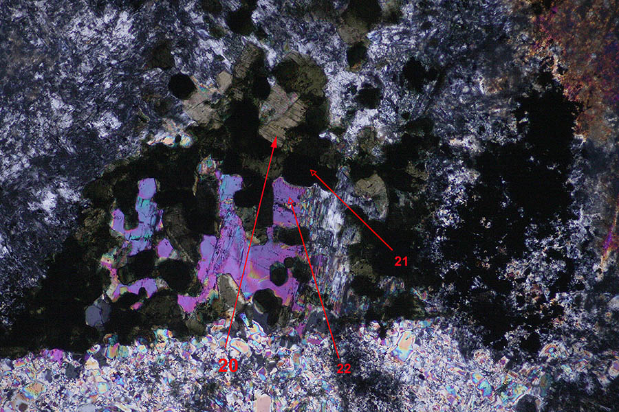
The green material labeled 20 on the picture above is the mica annite. Spectrum observed is quite similar to region 5 described previously. The birefringent crystal marked 22 is cancrinite. |
|
|
|
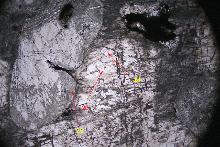
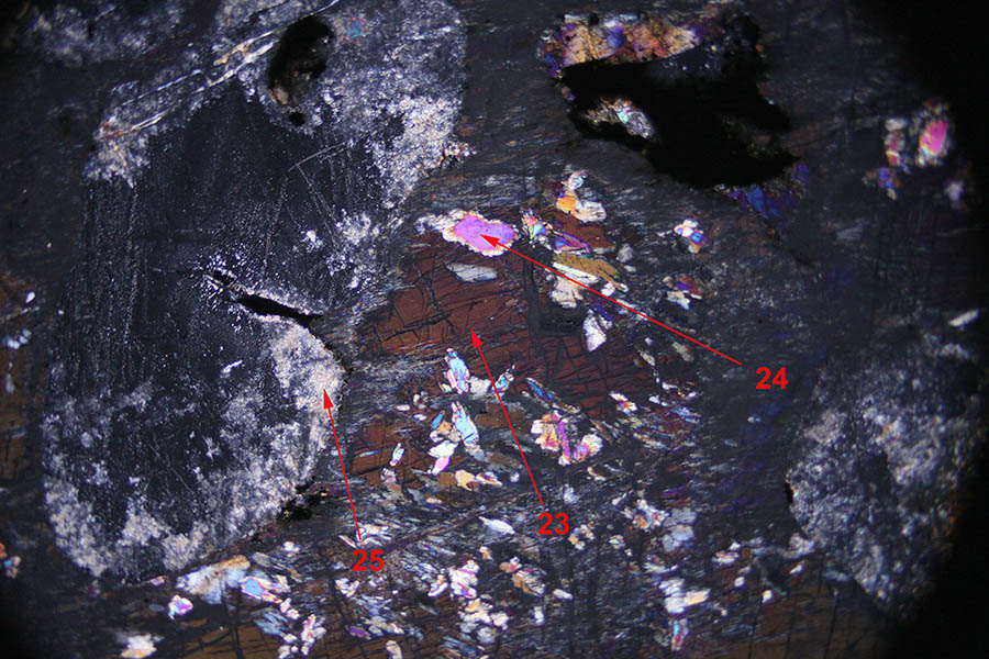
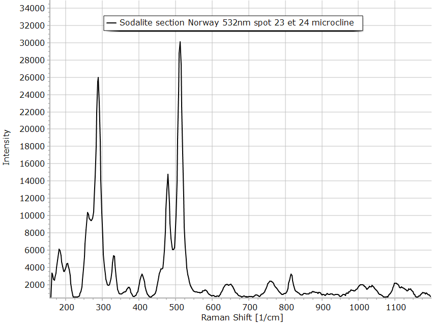
The right part of this figure is occupied by the feldspar microcline (23 and 24). On the left, the cubic sodalite particle is altered, the Raman spectrum of this alteration product is mesolite or natrolite. |
|
|

