Amphibolite from Ireland Raman spectra.
Thin section image of this rock.
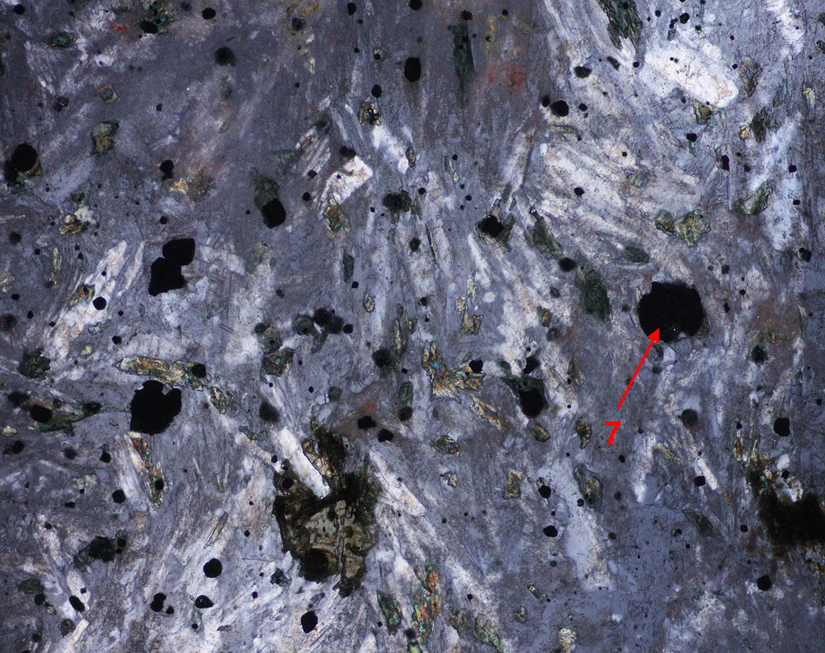
Amphibolite: amphiboles, biotite, quartz, dolomite, chlorite and magnetite.Locations of areas 1 to 3 in plane polarized view and cross polars image. |
|
 |
|
 |
|
 |
Areas 1 and 7 (images below) are amphiboles of the hornblende family with a pleochroism from green to yellow. The best match for the Raman spectra on the left is the sodium-calcium amphibole Taramite. |
 |
The brown pleochroic mineral (light brown to deep brown) is Biotite as shown in the spectrum on the left. Some of the biotite are cut perpendicularly to the acute bisectrix and thus appear very dark in the crossed polars view because the angle 2V between optic axes is very low. |
 |
The mineral of low birefringence in area 3 is evidently quartz as can be easily recognized in LPA view. |
Areas 4 and 5.Note that the brown biotite crystal at the top of the image is dark in the LPA view. |
|
 |
|
 |
|
 |
The high birefringence material in area 4 is dolomite. |
 |
The green crystal with very low birefringence in spot 5 (they appear very dark in LPA) is a material belonging to the chlorite family. |
Area 7: another example of the amphibole crystals.Raman spectrum 7 is reproduced in the first figure at the top of this page. |
|
 |
|
 |
|
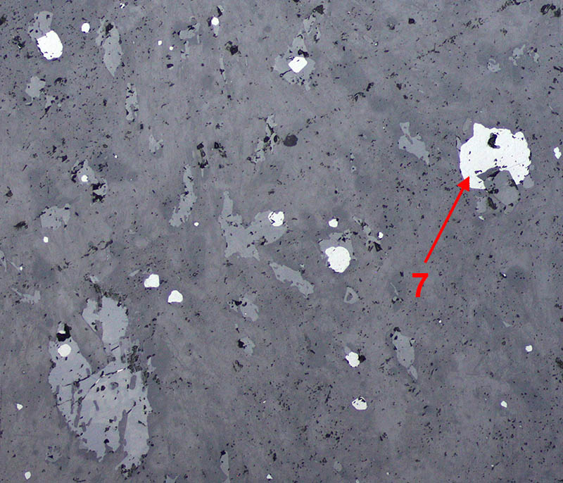
Opaques minerals in this section: magnetite. |
|
|
|
Reflection image in plane polarized
light: magnetite has a higher reflection coefficient than the silicates
around it. Below is the reflection image between crossed polars. |
 |
|
|
|
The blue curve on the left is the Raman spectrum obtained with an helium neon laser whose power has been reduced with a filter. The mineral is magnetite. Two small sharp peaks of an oxidation product are visible in the spectrum. If the full laser power is used, high additional peaks of oxidation can be seen. |

