Raman Weathered Peridotite from the Lanzo massive, Italy.
|
A thin section image of this rock can be seen here in plane polarized light and here in crossed polar view. The peridotite from Lanzo is heavily weathered in some part of the rock. Near the rock surface, no original mineral remains, all is transformed. This Raman study has been made with thick polished sections so the images of the location where Raman has been measured are reflected light pictures with or without crossed polarization.
The two images below illustrate the alteration of olivine crystals due to weathering. The original minerals are hydrated and transformed to other mineralogical materials, especially fine grained minerals. The end product of this transformation is serpentine, the low birefringence mineral seen just below on the crossed polars image. On the plane polarized micrograph, the serpentine is completely transparent without any structure due to its low refraction index. Between the remnant olivine crystals is a microcrystalline material, greenish in some places of the LPNA view, composed of a minerals mixture.
|


|
|
|
|
|
|
The areas 1, 2 and 3 exhibit increasingly refractive
index as can be seen on the left picture above: the reflection
coefficient is higher when the index of refraction increases. Thus we
expect three different minerals. On the double polarized image on the
right, the difference between regions 1 and 2 is not quite different.
This picture is an internal reflections image in a thick section so
material blow the surface is also imaged. The same is true for the Raman
microscopy. The microscope is not a confocal instrument so the thickness
being sampled is not absolutely restricted to the surface. The spectrum of the area 1 is mainly clinochlore but a small signal of olivine is also visible. It is a characteristic of this section: most spectra are not coming from a pure mineral but from a mineral assemblage due to the weathering. Area 2 is antigorite the serpentine material end member of the olivine alteration. Location 3, the blue curve, is the forsterite (olivine) as can be seen in comparison with the RRUFF reference material. The spectrum indicates the presence of the antigorite close to the high refraction index olivine. |
|
 |
||
|
|
|
|
|
|
The image above illustrates the transformation of a big
crystal to a microcrystalline material. On the plane polarized image,
the homogenous gray crystal (area 4) is transformed to a granular
material (area 5). On the crossed polars micrograph, The green
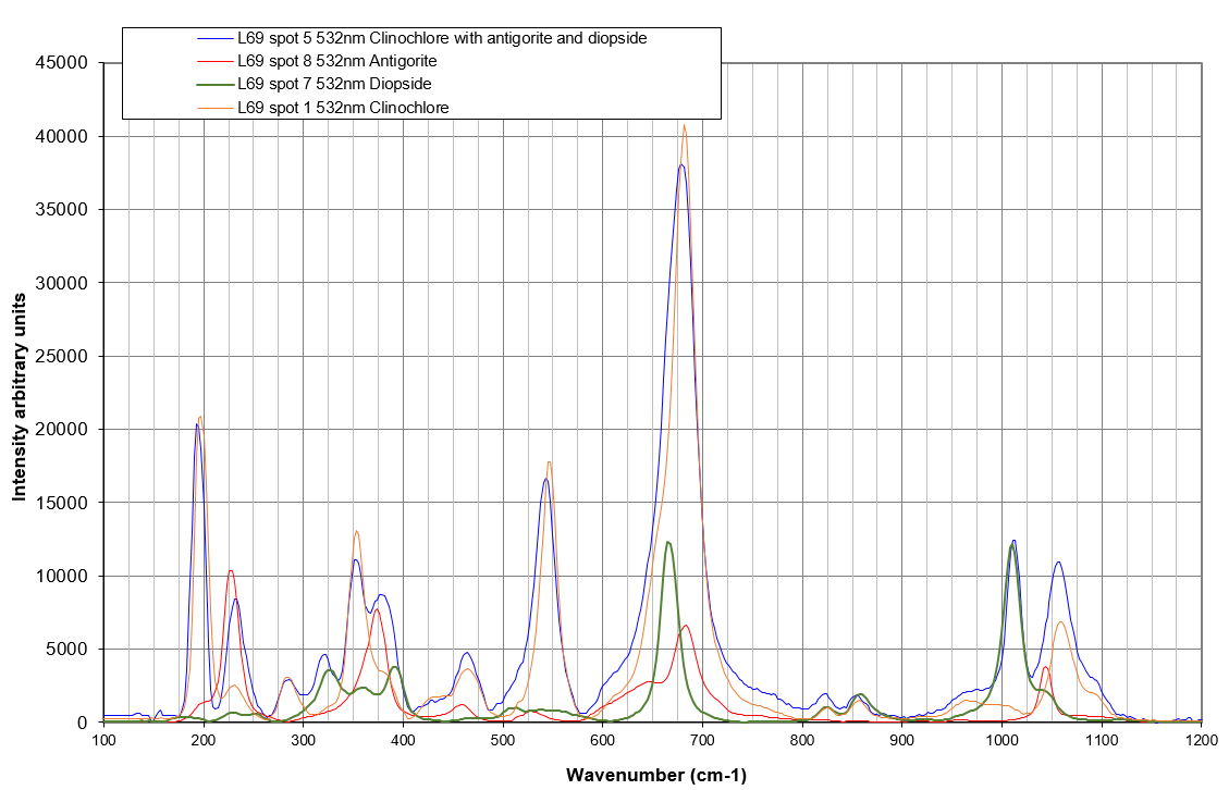
transparent crystal becomes a grainy white crystals mixture. The crystal labeled 4 is diopside as seen on the Raman spectrum on the left. The area 5 is a mineral mixture of diopside, antigorite and clinochlore. The pure minerals spectra taken at different locations have been added to the figure below for an easy comparison. |
|
|
|
||
|
|
 |
|
|
|
Different color as seen in the image above is not always synonymous of different minerals. The big crystal with cleavage planes (6) has the same index of refraction as the pale green crystal (7) as seen on the left image and the composition of both areas are the same: diopside. | |
|
|
|
|
|
|
|
|
|
|
Some other areas (8, 9, 10 and 11) have also been investigated with the Raman. The same minerals pure of in mixtures are found. Low reflectance in LPNA images is mainly clinochore, intermediate index is mainly antigorite and high index has a composition closer to olivine. | |
 |
In this section, we can also find some opaque minerals like magnetite and chromite as seen on the pictures and spectra included here. |
|
|
|
|
|


