Raman Apatite Chlorite section from Portugal.
A tin section image of this apatite - chlorite section can be found here.
Fluorescence images and spectra of this section are illustrated here.
 |
|
 |
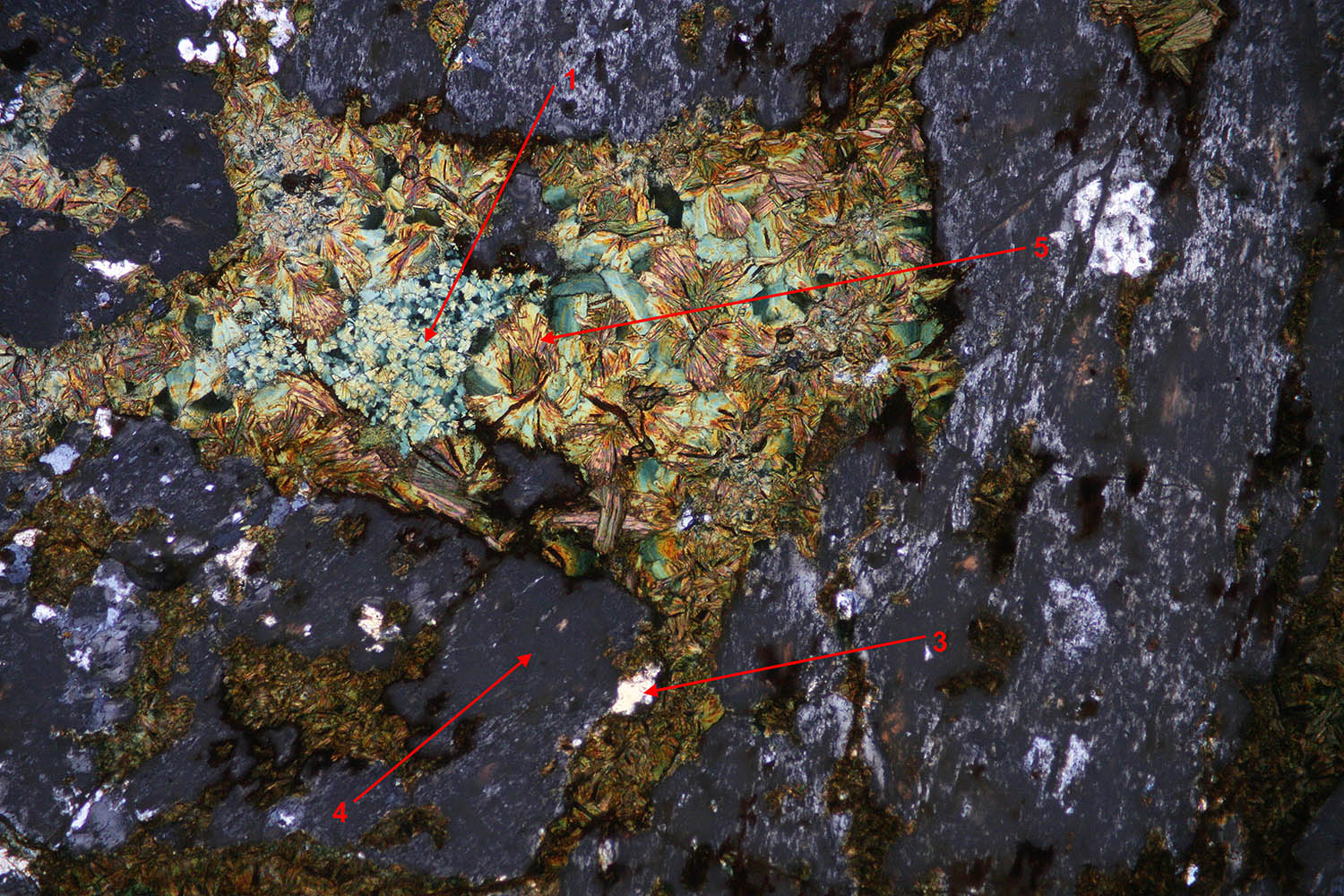
Image on top is a plane polarized general view of this
section with the 5 areas where Raman has been measured. The crystals 1 and 2, either green or white, are identified as chamosite, a member of the chlorite family. The spot 5 although exhibiting a different morphology has the same chamosite composition. Spectra are reproduced on the left. |
 |
|
 |
A few bright crystals like point 3 in the picture above (crossed polarizer and analyzer) are visible in this section. The spectrum on the left indicates the presence of quartz. |
 |
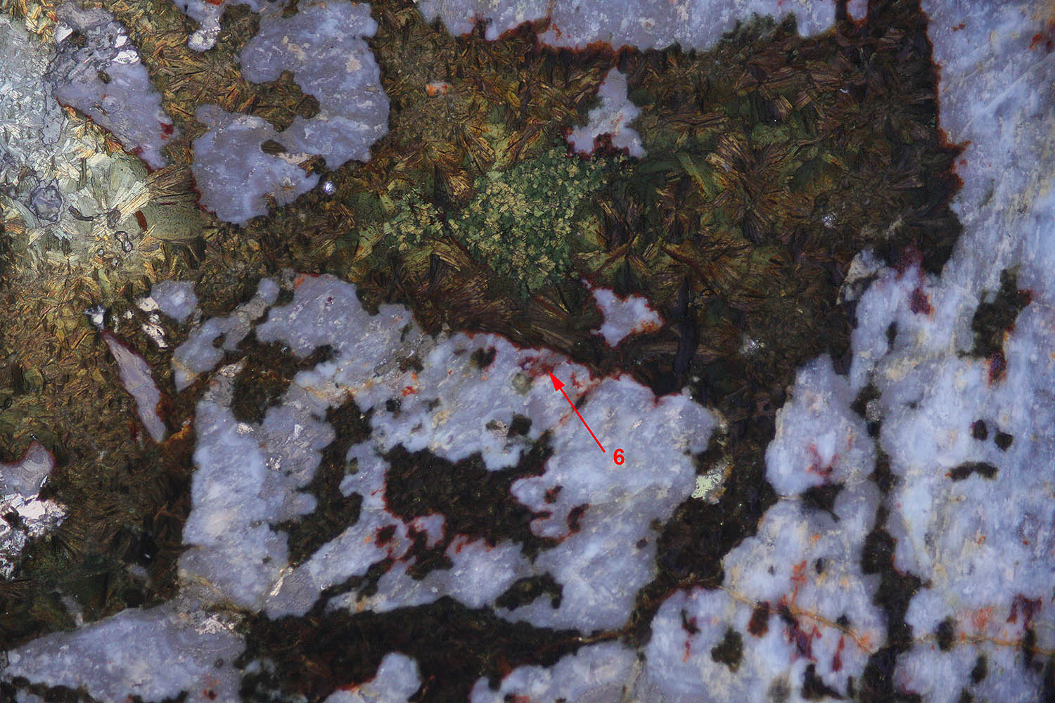
The area 4 is an altered material. The Raman spectroscopy identifies apatite in this region. The spectrum obtained from this area is not a good quality Raman spectrum due to the high fluorescence of this material. Zone 4 in the figure is completely dark in the LPA image above which indicates the presence of a low birefringence mineral like apatite. An interesting image is reproduced below: it is a reflection micrograph between crossed polars. It exhibits the internal reflections inside the section. In this view, the apatite areas appear white due to the fact that they are microcrystalline. A reflection fluorescence image is also given at the bottom: the center of the apatite regions emits a blue fluorescence and the rim an orange light. |
 |
|
 |
|
 |
In the reflection image, a red brown rim appears around
the apatite areas at the border with the chamosite regions. The Raman
spectrum indicates the presence of a limonite material. This minerals is
also present in cracks and veins of the section. The limonite is not
fluorescent and cannot thus be responsible of the orange fluorescence at
the border of each apatite area. The orange fluorescence around the
apatite could thus be due to a different composition of the apatite at
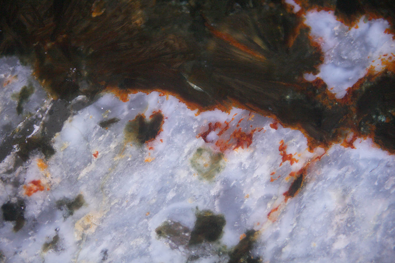
the contact of the chlorite. Below is a magnified image (200X) showing the structure of the red material. |
 |
|