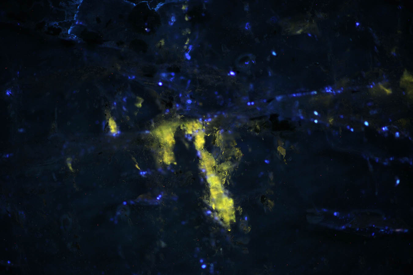
Fluorescence small inclusions in a Triplite section.
|
|
|
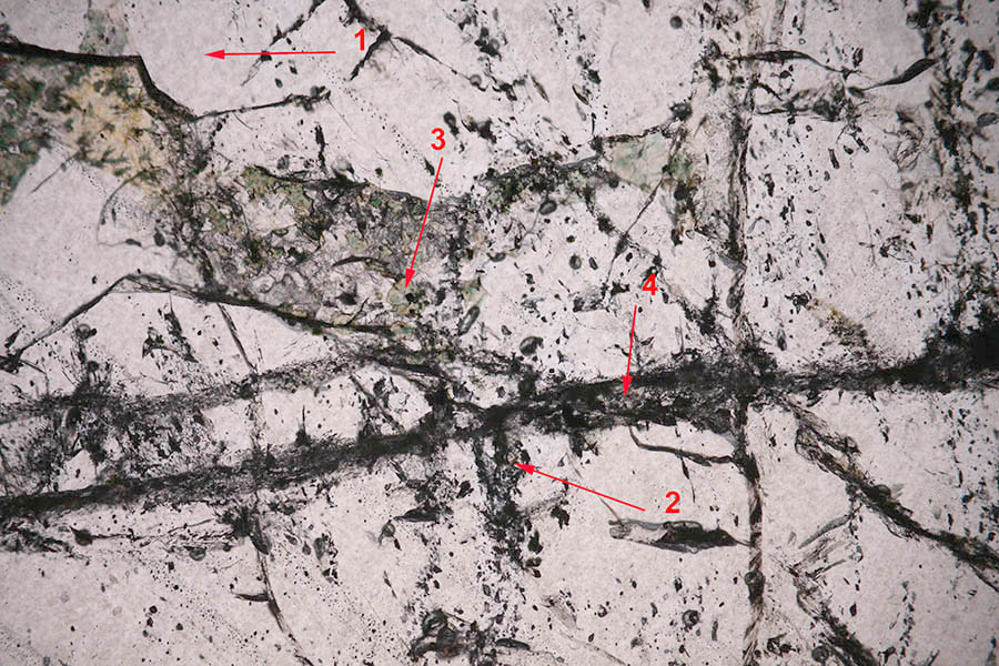
Fluorescent inclusions in triplite. The triplite occupies the whole section, it is not fluorescent, it appears thus very dark in this image.
|
|
|
|
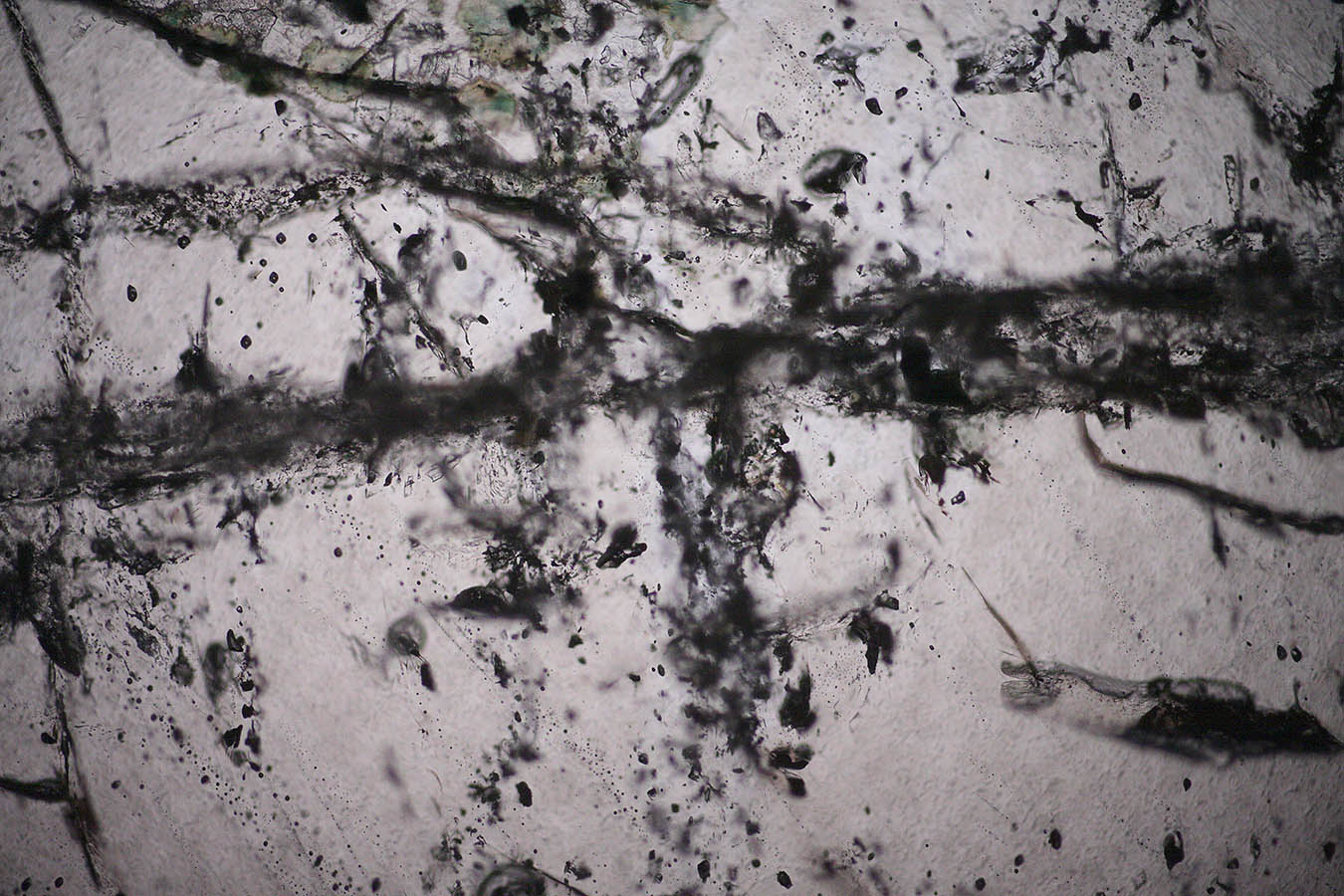
Same section in white light, transmission image. The fluorescent impurity is not clearly identified in this view.
|
|
|
|
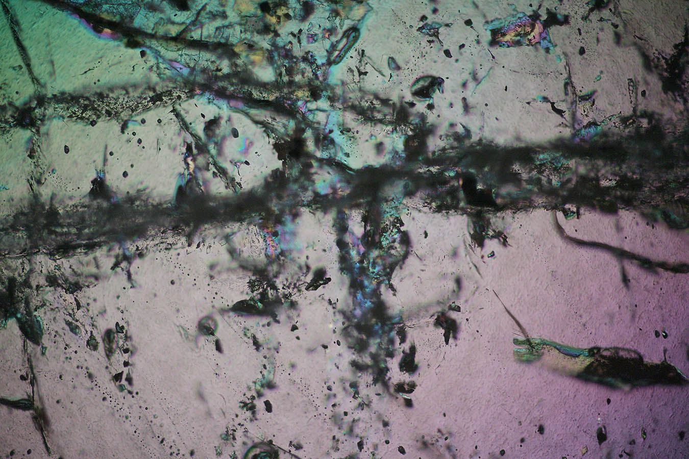
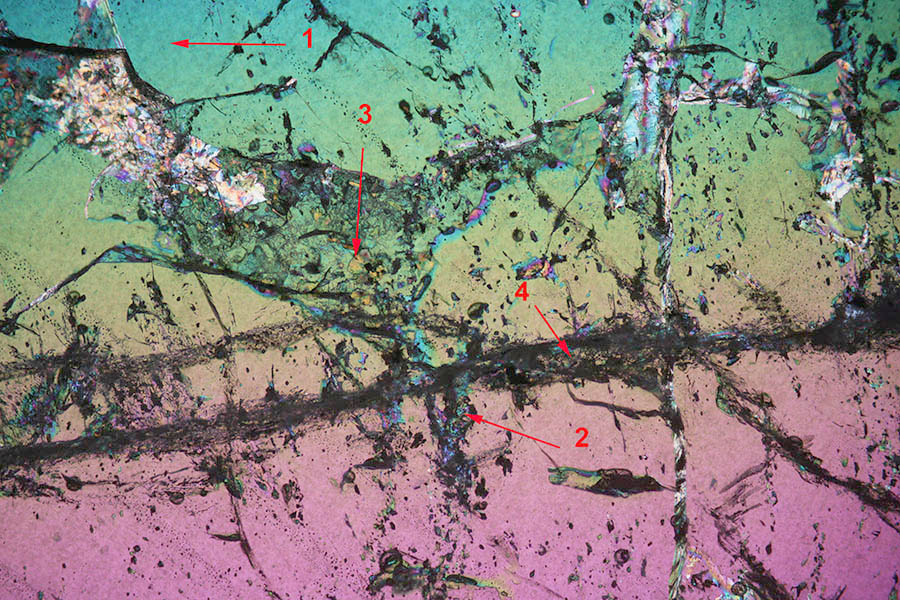
Crossed polars view of the same section.
|
|
|
|
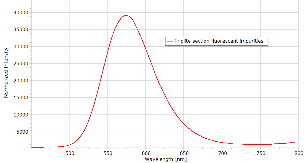
Spectrum of the fluorescent impurities in the triplite section.
|
 |
 |
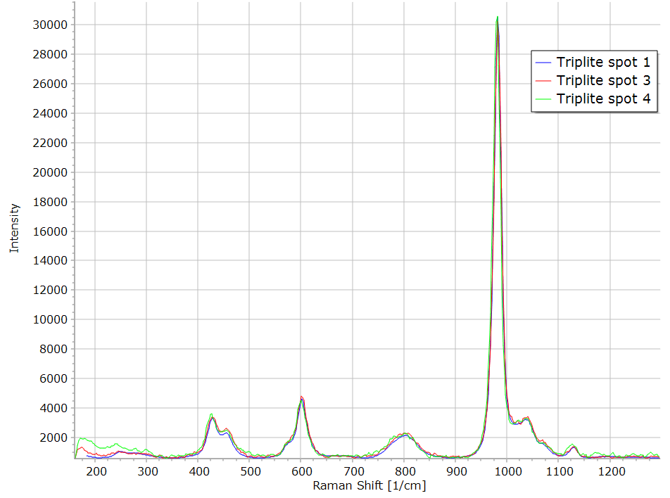 |
With the help of the Raman spectroscopy, I have tried to identify some impurities in the triplite section in particular in the fluorescence area. On the clear region number 1, a triplite spectrum is obtained. In the area 2, only a fluorescence signal can be obtained so no Raman identification has been possible. Green crystals in area 3 also give a triplite spectrum. They are quite easily identified on the transmission image but probably they are situated beneath the surface and surrounded by triplite so the Raman cannot identify them. In the fracture of the section (spot 4), again the triplite spectrum was found. |
 |
 |
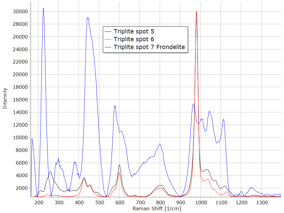 |
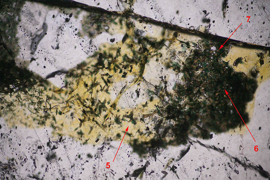
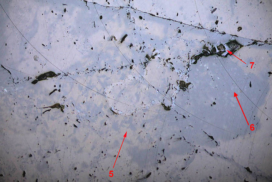
To identify the green brown microcrystals which appear in the triplite section, the reflection image could help. It gives an idea of the refraction index of the material situated at the surface. If the minerals present in the section are very transparent, the light is also reflected onto the bottom surface of the section (the glass plate) and it adds to the front reflected light. If some dark material is present in the section, as in area 6 for instance, a shadow of this material appears in the reflected light image. This phenomenon can be seen around zone 5. It is thus an artifact in the image, it does not mean that a lower refractive index material is present at the surface. If the reflected light is higher on the contrary, it means that another mineral could be present at the surface as observed in area 7. The spectrum obtained with the Raman in area 5 and 6 is again triplite. The spectrum 5 in the yellow region of the left figure has some difference with the normal triplite spectrum. On the other hand, the spectrum of mineral number 7, brighter in reflected light, is quite different, it is identified as frondelite a phosphate of Mn and Fe. |