Sodalite Fluorescence in Phalaborwa Rock Section.
Raman spectra of this section from Phalaborwa.
 |
|
Figure 1: fluorescent sodalite crystal. Transmission composite image, white light and 405 nm fluorescence image.
|
 |
|
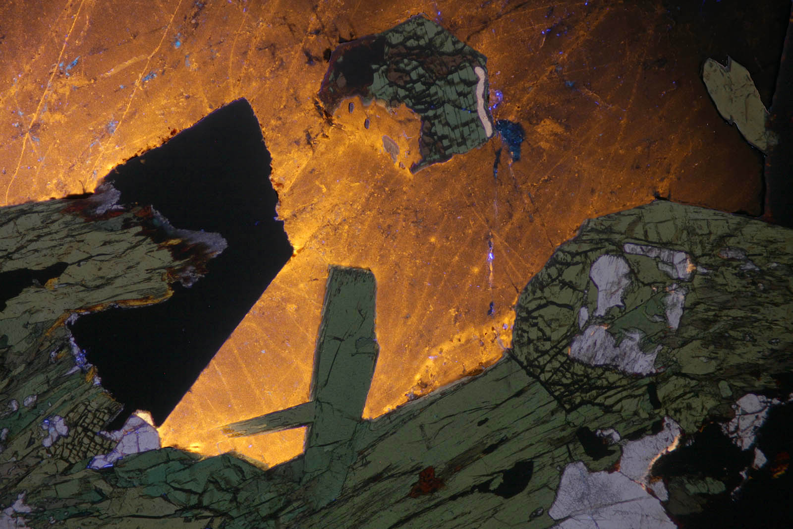
Figure 2: same section in plane polarized light. White sodalite, green diopside and aegirine.
|
|
|
|
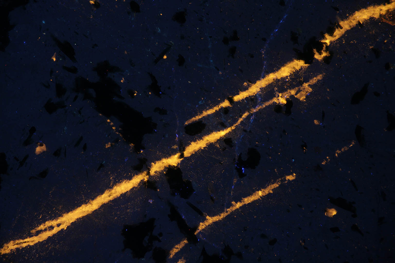
Figure 3: fluorescent veins of sodalite in nepheline.
|
 |
|
Figure 4:Composite image between LPA transmission image and 405 nm laser reflection image.
|
 |
|
Figure 5: plane polarized image of the same section. The sodalite is barely visible with this illumination, sodalite and nepheline are quite transparent.
|
 |
|
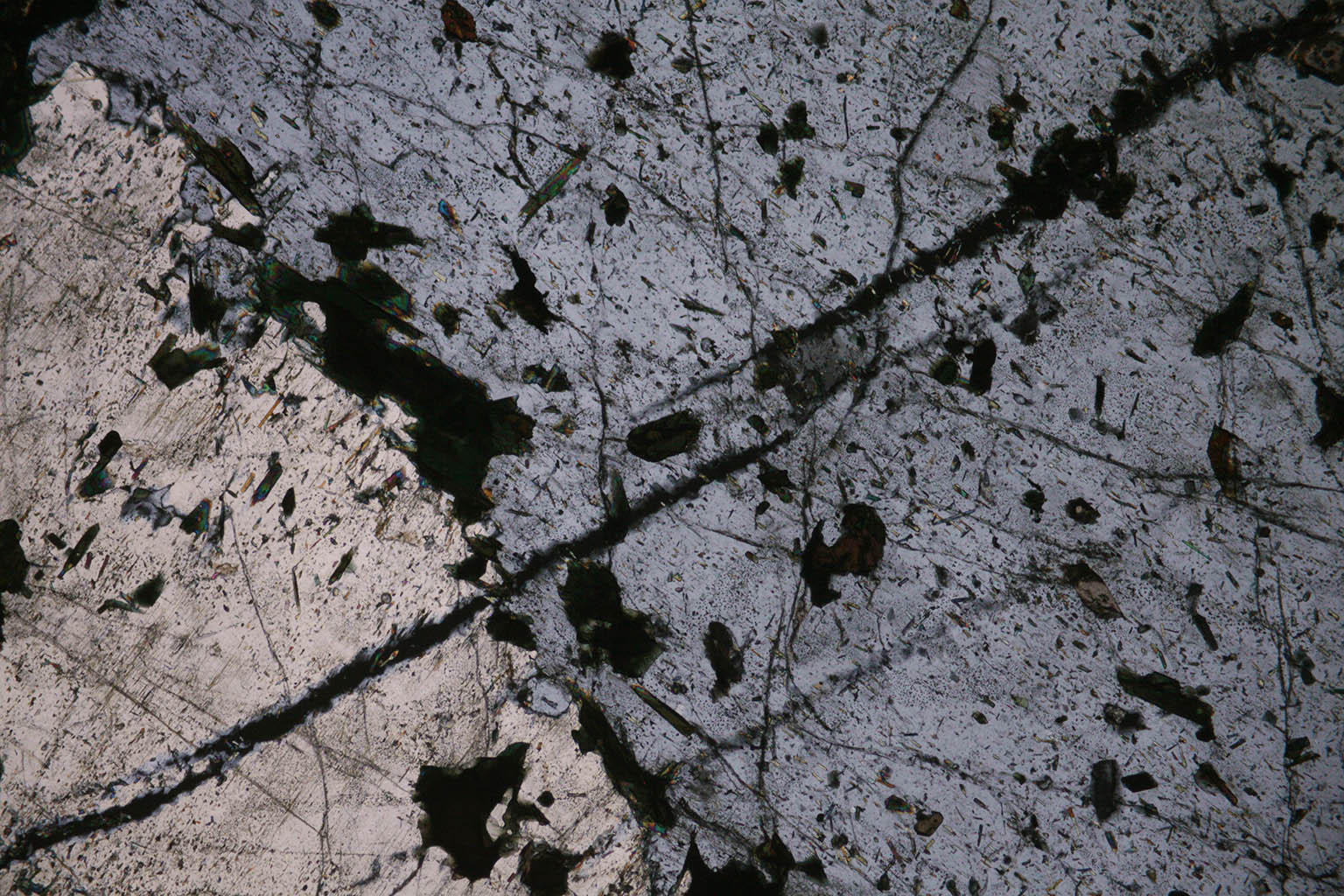
Figure 6: crossed polarizers view (LPA). The cubic sodalite appears black with this illumination. The birefringent nepheline is quite visible.
|
|
|
|
Fluorescence spectrum of the sodalite in Phalaborwa section.
|